
Registered Paramedical Training Center Details
Best Registered Paramedical Training Center. Mobile Phone 01797522136, 01987073965. HRTD Medical Institute is the best Registered Paramedical Training Center in Mirpur, Dhaka, Bangladesh. We provide almost all paramedical courses. Our Paramedical Courses are Paramedical Course 1 Year, Paramedical Course 2 Years, Paramedical Course 3 Years, Paramedical Course 4 Years, Diploma in Paramedical 2 Years, Diploma in Paramedical 3 Years, and Diploam in Paramedical 4 Years.

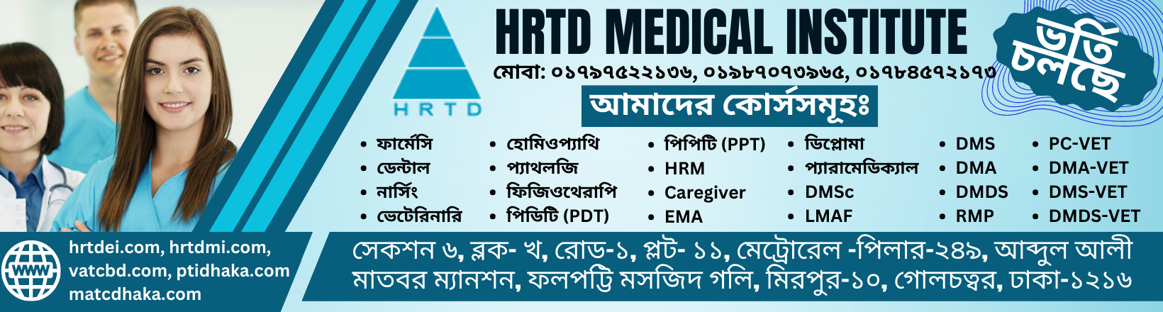
Location Address of Registered Paramedical Training Center
Location Address of Registered Paramedical Training Center. Mobile Phone 01797522136, 01987073965. HRTD Medical Institute, Abdul Ali Madbor Mansion, Section 6, Block-Kha, Raod-1, Plot 11, Metro Rail Piller No. 249, Mirpur 10 Golchattar, Dhaka-1216.
Teachers of Registered Paramedical Training Center
Dr. Sakulur Rahman, MBBS, CCD
Dr. Suhana, MBBS, PGT
Dr. Shamima, MBBS, PGT
Dr. Tisha, MBBS, PGT
Dr. Disha, MBBS
Dr. Lamia, MBBS
Dr. Laila, MBBS
Dr. Farabi, MBBS
Dr. Turzo, MBBS
Dr. Sanjana, BDS,
Dr. Juthi, BDS
Dr. Keya, BDS
Dr. Nurunnahar, BDS
Dr. Rezoan, MBBS
Dr. Mahinul, MBBS
Subjects for Paramedical Courses of Registered Paramedical Training Center
Human Anatomy and Physiology
Pharmacology-1
First Aid
Study of OTC Drugs
Hematology
General Pathology
Pathology for Medical Practice
Microbiology
Antimicrobial Drugs
Cardiovascular Anatomy & Physiology
Cardiovascular Drugs & Medicine
Gastro Anatomy & Physiology
Gastrointestinal Drugs & Medicine
Gastrology
Orthopedic Anatomy & Physiology
Orthopedic Drugs & Medicine
Common Orthopedic Diseases
Neuro Anatomy & Physiology
Neurological Drugs & Medicine
Medicine Diagnosis
Histology & Cytology
Medical Embriology
Practice of Medicine-1
Neuromedicine
Orthopedic Disease & Treatment
Common Dermatological Diseases
Ophthalmic Drugs & Medicine
ENT Drugs & Medicine
Mental Health & Psychotherapy
Common Infectious Disease
Human Anatomy and Physiology for Paramedical Courses
The study of the Body Structure and its functions is Anatomy and Physiology. Here we discuss the systems of the Human Body and its Organs, Tissues, and Cells. The systems of the Human Body are the Digestive System, Respiratory System, Cardiovascular System, Skeletal System, Muscular System, Nervous System, Endocrine System, Immune System, Lymphatic System, Integumentary System, and Urinary System.
Foundational concepts
- Levels of organization: The human body is organized hierarchically, from atoms and molecules to cells, tissues, organs, and organ systems.
- Homeostasis: The body’s ability to maintain a stable internal environment, which is crucial for survival. This includes regulating factors like temperature, blood pressure, and nutrient levels.
- Structure and function: A fundamental principle is that a body part’s structure is intricately related to its function.
Major body systems
- Integumentary system: The skin, hair, and nails, which act as the body’s protective outer covering.
- Skeletal system: Bones, joints, and cartilage that provide support and structure for the body.
- Muscular system: Muscles that enable movement through contraction.
- Nervous system: The brain, spinal cord, and nerves that coordinate and control body functions, including sensing and movement.
- Cardiovascular system: The heart, blood vessels, and blood that transport oxygen, nutrients, and waste throughout the body.
- Respiratory system: Structures like the lungs, which facilitate gas exchange (oxygen in, carbon dioxide out).
- Endocrine system: Glands that produce hormones to regulate body processes.
- Digestive system: Processes that break down food and absorb nutrients.
- Urinary system: Kidneys and other organs that filter waste from the blood.
- Reproductive system: Organs related to reproduction.
- Immune system: Defends the body against pathogens and diseases.
Areas of study
- Anatomy: The study of body structures, which can be seen with the naked eye (gross anatomy) or require a microscope (microscopic anatomy).
- Physiology: The study of how the body’s structures and systems function.
- Cellular and tissue-level study: This includes cytology (study of cells) and histology (study of tissues).
Digestive System :
The main topics of the digestive system’s anatomy and physiology are the GI tract organs (mouth, esophagus, stomach, small intestine, large intestine, anus), accessory organs (liver, pancreas, gallbladder, salivary glands), and the six main functions: ingestion, propulsion, mechanical and chemical digestion, absorption, and elimination. Key physiological processes include peristalsis (muscle contractions to move food), enzyme secretion, and the breakdown of food into absorbable nutrients.
Anatomy
- Gastrointestinal (GI) Tract: A hollow, muscular tube that runs from the mouth to the anus.
- Mouth: Where food is ingested and mechanical and chemical digestion begins.
- Esophagus: A tube that transports food from the pharynx to the stomach.
- Stomach: A muscular bag that mixes food with acid and enzymes to create a paste called chyme.
- Small Intestine: The primary site for nutrient absorption, consisting of the duodenum, jejunum, and ileum.
- Large Intestine: Absorbs water and electrolytes, and includes the cecum, colon, rectum, and appendix.
- Rectum and Anus: Store and eliminate waste, respectively.
- Accessory Organs: Organs that aid digestion by secreting substances into the GI tract.
- Salivary Glands: Produce saliva to help moisten food and begin carbohydrate digestion.
- Liver: Produces bile to help digest fats.
- Gallbladder: Stores and concentrates bile.
- Pancreas: Produces digestive enzymes and bicarbonate to neutralize stomach acid.
Physiology
- Ingestion: Taking food into the body through the mouth.
- Propulsion: The movement of food through the GI tract, mainly through peristalsis.
- Mechanical Digestion: The physical breakdown of food through chewing, churning, and mixing.
- Chemical Digestion: The use of enzymes and acids to break down food into smaller molecules.
- Absorption: The process of moving nutrients from the digestive tract into the bloodstream or lymphatic system.
- Elimination: The expulsion of indigestible waste products from the body as feces.
Respiratory System
The main topics of respiratory system anatomy and physiology include its primary function of gas exchange (oxygen in, carbon dioxide out), the structures of the upper and lower respiratory tracts (nose, pharynx, larynx, trachea, bronchi, and lungs/alveoli), and the mechanics of breathing driven by the diaphragm and intercostal muscles. Key physiological processes include warming/humidifying air, conducting it to the lungs, and gas exchange in the alveoli.
Anatomy: structures of the respiratory system
- Upper Respiratory Tract:
- Nose and nasal cavity: Warms, humidifies, and filters incoming air.
- Pharynx (throat): Common passageway for air and food.
- Larynx (voice box): Contains the vocal cords and prevents food from entering the airway.
- Lower Respiratory Tract:
- Trachea (windpipe): Carries air to the bronchi; supported by C-shaped cartilage rings.
- Bronchi: The trachea divides into the right and left main bronchi, which branch into smaller bronchi and bronchioles.
- Lungs: The main organs, divided into lobes (right has three, left has two) and protected by the rib cage.
- Alveoli: Tiny air sacs at the end of the bronchioles where gas exchange occurs.
Physiology: functions of the respiratory system
- Gas Exchange: The primary function is to take in oxygen and expel carbon dioxide. Oxygen from the alveoli diffuses into the blood, and carbon dioxide diffuses from the blood into the alveoli to be exhaled.
- Breathing Mechanics (Ventilation):
- Inhalation: The diaphragm and intercostal muscles contract, increasing the volume of the thoracic cavity and drawing air into the lungs.
- Exhalation: These muscles relax, decreasing the volume and pushing air out of the lungs.
- Air Conditioning: The respiratory system warms, humidifies, and filters the air as it passes through the nasal cavity and airways.
- Protection: Cilia and mucus in the airways trap and remove debris (mucociliary escalator), and the epiglottis prevents food from entering the trachea.
- Other functions: The larynx is essential for voice production.
Cardiovascular System
The main topics of the cardiovascular system are its two main anatomical components, the heart and the blood vessels (arteries, veins, and capillaries), which work together to transport blood. Key physiological aspects include the two main circulatory circuits (pulmonary and systemic), the electrical conduction system that controls heart rate, the cardiac cycle of contraction and relaxation, and the regulatory mechanisms that maintain adequate blood flow to meet the body’s needs.
Anatomy
- The Heart: A four-chambered muscular pump with two atria (receiving chambers) and two ventricles (pumping chambers). The septum divides the right and left sides.
- Blood Vessels: A network of tubes that carry blood throughout the body.
- Arteries: Carry blood away from the heart.
- Veins: Carry blood back to the heart.
- Capillaries: Tiny vessels that connect arteries and veins, where exchange of gases and nutrients occurs.
- Heart Valves: Structures that ensure blood flows in only one direction through the heart.
- Heart Wall: Composed of three layers: the epicardium (outer), myocardium (muscular middle), and endocardium (inner).
Physiology
- Circulatory Circuits:
- Pulmonary Circuit: Carries deoxygenated blood from the heart to the lungs and returns oxygenated blood to the heart.
- Systemic Circuit: Transports oxygenated blood from the heart to the rest of the body and returns deoxygenated blood to the heart.
- Coronary Circuit: Supplies the heart muscle itself with blood.
- Cardiac Cycle: The sequence of events in one complete heartbeat, consisting of two main phases:
- Diastole: Relaxation and filling of the ventricles.
- Systole: Contraction and pumping of blood out of the heart.
- Electrical Conduction System: A system that generates and conducts electrical impulses to coordinate heart contractions.
- Sinoatrial (SA) Node: The heart’s natural pacemaker.
- Atrioventricular (AV) Node, Bundle of His, Purkinje Fibers: A pathway that spreads the electrical signal to the ventricles.
- Regulation: The cardiovascular system is regulated to ensure blood flow meets the body’s needs.
- Autonomic Nervous System: Sympathetic and parasympathetic nerves influence heart rate and blood vessel contraction.
- Endocrine System: Hormones affect blood flow and heart function.
- Autoregulatory Mechanisms: Local factors help control blood flow to different tissues.
Muscular System
The main topics in the anatomy and physiology of the muscular system include the three types of muscle tissue (skeletal, smooth, and cardiac), the physiology of muscle contraction involving actin and myosin, the structure and function of the muscular system including its role in movement, posture, and heat generation, and the supportive structures like tendons and fascia.
Types of muscle tissue
- Skeletal muscles: Voluntary muscles attached to bones that are responsible for movement, posture, and generating heat.
- Smooth muscles: Involuntary muscles found in the walls of hollow organs like the digestive and urinary tracts, and blood vessels. They propel substances through these organs.
- Cardiac muscle: Involuntary muscle found only in the heart, responsible for pumping blood throughout the body.
Anatomy and structure
- Muscle composition: A single muscle is made up of many tissues, including blood vessels, nerves, and muscle fibers.
- Connective tissue: Connective tissue layers like the epimysium, perimysium, and endomysium organize muscle fibers into fascicles and then into the whole muscle.
- Muscle fiber structure: Within each muscle fiber are myofibrils, which contain contractile proteins called actin and myosin.
- Sarcomeres: The functional units of a muscle fiber are sarcomeres, defined by Z-lines. The sliding of actin and myosin past each other within the sarcomere is what causes muscle contraction.
- Supportive structures:
Physiology and function
- Muscle contraction: An electrical signal from a motor neuron triggers the release of a chemical messenger, which causes the actin and myosin filaments to slide past one another, shortening the sarcomere and contracting the muscle.
- Movement and posture: Muscles are the primary movers, working in groups to create movement, maintain posture, and stabilize joints.
- Heat generation: Muscle contractions generate most of the body’s heat, helping to maintain a stable body temperature. Shivering is an example of rapid, involuntary muscle contractions to generate heat.
- Organ protection: Muscles help protect internal organs.
- Functional roles:
- Prime movers (agonists): The primary muscle responsible for a specific movement.
- Antagonists: Oppose the action of prime movers to allow for more controlled movements.
- Synergists: Assist prime movers by adding force or reducing undesirable movements.
Skeletal System
The main topics in skeletal system anatomy and physiology include the system’s functions (support, movement, protection, blood cell production, mineral storage), its two divisions (axial and appendicular skeletons), bone tissue composition and remodeling, and different types of joints and their movements. Key components are bones, cartilage, and ligaments, with adult humans having approximately 206 bones.
1. Functions of the skeletal system
- Support: Provides a rigid framework for the body and a point of attachment for muscles and other tissues.
- Movement: Acts as a system of levers for muscles, enabling body movement at joints.
- Protection: Encloses and protects vital internal organs, such as the brain (cranium), heart and lungs (rib cage), and spinal cord (vertebrae).
- Blood cell production: Produces red blood cells, white blood cells, and platelets in the red bone marrow found within certain bones.
- Mineral storage: Stores and releases essential minerals like calcium and phosphorus, which can be released into the bloodstream when needed.
- Energy storage: Stores fat in the yellow bone marrow.
2. Divisions of the skeleton
- Axial skeleton: The central axis of the body, which includes the skull, vertebral column, and ribcage.
- Appendicular skeleton: The framework of the limbs and girdles, including the shoulder girdle, pelvic girdle, arms, and legs.
3. Bone tissue and remodeling
- Composition: Bone is a living connective tissue made of cells (osteoblasts, osteocytes, osteoclasts) and a matrix of collagen and mineral salts. Cartilage, another connective tissue, provides cushioning in joints.
- Bone remodeling: The continuous process of old bone tissue being broken down and new bone tissue being formed, which can be influenced by factors like exercise and hormones.
4. Types of joints
- Ball-and-socket: Allows for multi-axial movement (e.g., hip and shoulder).
- Hinge: Permits movement in one plane, like flexion and extension (e.g., knee and elbow).
- Pivot: Enables rotation (e.g., the joint between the first two vertebrae in the neck).
- Plane: Allows for gliding movements (e.g., between the bones of the wrist and ankle).
- Saddle: Provides biaxial movement (e.g., the joint at the base of the thumb).
- Condyloid: Allows for movement in two different planes (e.g., the wrist joint).
Nervous System
The main topics of the nervous system’s anatomy and physiology are its division into the Central Nervous System (CNS) (brain and spinal cord) and the Peripheral Nervous System (PNS) (nerves and ganglia). The CNS processes information, while the PNS transmits signals between the CNS and the rest of the body. The PNS is further divided into the Somatic Nervous System (voluntary movement) and the Autonomic Nervous System (involuntary functions), which includes the sympathetic (“fight or flight”) and parasympathetic (“rest and digest”) systems.
Central Nervous System (CNS)
- Brain: The control center for thought, memory, movement, and bodily functions. It is protected by the skull, meninges, and cerebrospinal fluid. Key areas include:
- Cerebrum: The largest part of the brain, responsible for higher-level functions like thought, learning, and memory.
- Cerebellum: Coordinates movement and balance.
- Brainstem: Connects the cerebrum and cerebellum to the spinal cord and controls vital functions like breathing and heart rate.
- Hypothalamus and Thalamus: Located deep within the brain, the hypothalamus regulates body temperature, thirst, and hunger, while the thalamus acts as a relay center for sensory and motor signals.
- Spinal Cord: A long, column-like structure that extends from the brainstem and is protected by the vertebrae. It serves as the main pathway for signals between the brain and the rest of the body and also handles some reflexes independently.
Peripheral Nervous System (PNS)
- Structure: Comprises all the nerves that branch out from the CNS. These nerves are bundles of axons (nerve fibers) and are covered by layers of connective tissue.
- Somatic Nervous System: Controls all voluntary movements, such as walking or picking up objects, by sending signals to skeletal muscles.
- Autonomic Nervous System: Manages involuntary functions that the body performs automatically, like breathing, digestion, and heart rate. It has two main divisions:
- Sympathetic: Activates the “fight or flight” response, preparing the body for action.
- Parasympathetic: Promotes the “rest and digest” response, calming the body and returning it to a state of equilibrium.
Fundamental Unit
- Neuron: The basic structural and functional unit of the nervous system, responsible for transmitting electrochemical signals.
- Neuroglia: Supporting cells that insulate, protect, and provide nourishment to neurons. Examples include astrocytes, microglia, oligodendrocytes, and Schwann cells.
Endocrine System
The main topics in the anatomy and physiology of the endocrine system include its overall function, the major glands and their locations, the hormones they produce, and the regulation of the system. The system regulates vital processes like growth, metabolism, reproduction, and homeostasis through chemical messengers called hormones, which are secreted by glands like the hypothalamus, pituitary, thyroid, adrenals, pancreas, and gonads.
Anatomy: Major endocrine glands and organs
- Hypothalamus: Located at the base of the brain, it links the nervous system to the endocrine system via the pituitary gland.
- Pituitary gland: Often called the “master gland,” it receives signals from the hypothalamus and controls many other endocrine glands.
- Thyroid and parathyroid glands: Located in the neck, the thyroid regulates metabolism, while the parathyroids manage blood calcium levels.
- Adrenal glands: Situated on top of the kidneys, they produce hormones like cortisol and epinephrine that manage stress, blood pressure, and metabolism.
- Pancreas: A gland with both endocrine (islets of Langerhans) and exocrine functions, it secretes insulin and glucagon to regulate blood sugar.
- Gonads: These include the ovaries (in females) and testes (in males), which produce sex hormones and germ cells.
- Pineal gland: Located in the brain, it produces melatonin to regulate sleep cycles.
Physiology: Functions and processes
- Hormone production and secretion: Endocrine glands secrete hormones directly into the bloodstream to act as chemical messengers, targeting specific cells throughout the body.
- Homeostasis: The system maintains a stable internal balance for processes like blood sugar (insulin, glucagon), blood calcium (parathyroid hormone), and water balance.
- Regulation: The hypothalamus and pituitary gland form a control center that regulates other glands. For example, the hypothalamus stimulates the pituitary to release hormones that control the thyroid, adrenals, and gonads.
- Growth and metabolism: The endocrine system is crucial for tissue growth, maturation, and regulating metabolism to maintain body temperature and mental function.
- Reproduction: It controls the development and function of reproductive systems and regulates processes like puberty, pregnancy, and childbirth.
- Other functions: The system also manages the stress response (“fight-or-flight”), heart rate and blood pressure, immune responses, and nutrient levels.
Reproductive System
The main topics in the anatomy and physiology of the reproductive system include the gonads (testes and ovaries), which produce gametes (sperm and eggs) and hormones; the internal and external genitalia (ducts, glands, and organs like the uterus, prostate, and penis); and the hormonal regulation and cycles that govern sexual development and function. These structures work together to facilitate reproduction by producing and transporting gametes, enabling fertilization, and, in females, supporting a developing fetus.
Male reproductive system
- Gonads: The testes produce sperm and the male sex hormone, testosterone.
- Internal and external genitalia:
- Epididymis: Where sperm mature and are stored.
- Vas deferens: The tube that transports sperm from the epididymis.
- Accessory glands: Seminal vesicles, the prostate, and the bulbourethral glands produce the fluid portion of semen.
- Urethra: The tube that carries semen and urine out of the body.
- Penis: The external organ used for sexual intercourse.
- Hormonal regulation: Testosterone is crucial for sperm production, secondary sex characteristics, and libido.
Female reproductive system
- Gonads: The ovaries produce eggs (ova) and female sex hormones like estrogen and progesterone.
- Internal and external genitalia:
- Ovaries: Produce eggs.
- Fallopian tubes: Transport the egg from the ovary to the uterus.
- Uterus: Where a fertilized egg implants and a fetus develops.
- Vagina: Connects the uterus to the outside of the body.
- Vulva: The external female genitalia, including the labia.
- Hormonal regulation: The menstrual cycle, regulated by estrogen and progesterone, includes the development of an egg (follicular phase), its release (ovulatory phase), and the preparation of the uterus for pregnancy.
Key functions and processes
- Gametogenesis: The creation of sperm (spermatogenesis) and eggs (oogenesis).
- Hormone production: Both systems rely on hormones to regulate development and function.
- Fertilization: The fusion of a sperm and an egg to form a zygote.
- Gestation: The period of pregnancy, in which the female’s body supports the developing fetus, often involving the formation of a placenta.
- Puberty: The period when the reproductive systems mature and become capable of reproduction.
Urinary System
The main topics in the anatomy and physiology of the urinary system include its components—kidneys, ureters, bladder, and urethra—and their functions, which involve filtering blood to produce urine, regulating water and pH balance, and eliminating waste from the body. The functional unit of the kidney is the nephron, where filtration, reabsorption, and secretion occur.
Anatomy of the urinary system
- Kidneys: Two bean-shaped organs located on either side of the spine that filter blood. Internally, they contain a cortex and medulla, which house millions of nephrons.
- Ureters: Muscular tubes that transport urine from the kidneys to the bladder.
- Bladder: A hollow, muscular sac that stores urine.
- Urethra: A tube that connects the bladder to the outside of the body, allowing urine to be expelled.
Physiology of the urinary system
- Filtration: Blood is filtered in the glomerulus, a network of capillaries within the nephron, to remove waste products, excess water, and salts.
- Reabsorption and Secretion: As the filtrate passes through the renal tubule, essential substances like water and glucose are reabsorbed back into the blood, while additional waste products and ions are secreted into the tubule.
- Hormonal Regulation: The kidneys produce hormones that regulate blood pressure (renin) and stimulate red blood cell production (erythropoietin).
- Fluid and Electrolyte Balance: The system regulates the body’s water and electrolyte balance, including sodium and potassium levels.
- Acid-Base Balance: The urinary system plays a role in maintaining the body’s long-term acid-base balance.
- Urine Elimination: When the bladder fills, stretch receptors trigger the micturition reflex. The brain can then control the voluntary relaxation of the external sphincter, allowing urine to exit the body.
Immune System
The main topics of the urinary system are its organs (kidneys, ureters, bladder, urethra), its function (filtering blood to produce and expel urine, which removes waste), and common issues (like infections and stones).
Organs and their functions
- Kidneys: The primary organs that filter waste and excess fluid from the blood to produce urine. They also regulate blood pressure, pH, and water volume.
- Ureters: Tubes that transport urine from the kidneys to the bladder.
- Bladder: A hollow organ that stores urine until it’s ready to be expelled from the body.
- Urethra: The tube that carries urine from the bladder out of the body during urination.
Key functions
- Waste removal: The system filters metabolic waste products like urea, uric acid, and excess salts from the bloodstream.
- Fluid and electrolyte balance: It helps regulate the body’s water balance and the concentration of electrolytes like sodium and potassium.
- Blood pressure regulation: The kidneys play a significant role in controlling blood pressure.
- pH regulation: It helps maintain the body’s acid-base balance.
Common problems
- Urinary tract infections (UTIs): Infections that often affect the bladder and can spread to the kidneys.
- Kidney stones: Solid masses that form in the kidneys from minerals in the urine.
- Incontinence: The loss of bladder control.
- Enlarged prostate: In men, this can make it difficult to urinate by pressing on the urethra.
Lymphathic System
The main topics of the lymphatic system are its structure (a network of vessels, nodes, and organs), its functions (fluid balance, fat absorption, and immune defense), and its role in the immune response. It works like a secondary circulatory system, collecting excess fluid from tissues, filtering it for waste and pathogens in the lymph nodes, and returning it to the blood. It also absorbs fats from the intestines and contains immune cells to fight infection.
Structure
- Lymphatic vessels: A network of vessels that collect and transport lymph, similar to blood vessels.
- Lymph nodes: Small glands that filter lymph and store immune cells, which is why they can swell when you’re sick.
- Lymphoid organs: Include the spleen (filters blood), tonsils (trap pathogens), and thymus (site of T-cell development).
- Lymph: A fluid that circulates through the vessels, containing white blood cells, proteins, and fats.
Functions
- Fluid balance: Drains excess fluid from tissues, preventing swelling and maintaining blood volume.
- Fat absorption: Absorbs fats and fat-soluble vitamins from the digestive system and transports them to the blood.
- Immune defense: Filters out waste, bacteria, and cancer cells. It also serves as a highway for immune cells to travel throughout the body.
Immune Response
- Filtering: Lymph nodes act as checkpoints where immune cells can detect and destroy pathogens in the lymph.
- Immune cell production: The primary lymphoid organs, like the bone marrow and thymus, are where white blood cells (lymphocytes) are produced and mature.
- Immune surveillance: Provides a site for immune cells to monitor the body for threats and mount an immune response.
Covering System
The main topics in the anatomy and physiology of the covering system (integumentary system) focus on its structure (skin layers and accessory organs) and its diverse functions (protection, temperature regulation, sensation, and more).
I. Anatomy of the Integumentary System
The integumentary system consists of the skin and its accessory structures.
A. Skin Layers
The skin is composed of three main layers:
- Epidermis: The outermost, avascular (no blood supply) layer made of stratified squamous epithelium. Its primary cells are keratinocytes, which produce the tough, waterproof protein keratin. Melanocytes, which produce the pigment melanin, are also located here.
- Dermis: The middle layer composed of dense irregular and areolar connective tissue. It contains blood vessels, nerves, hair follicles, sweat glands, and oil glands. It has two regions: the superficial papillary layer and the deeper reticular layer, which gives the skin its elasticity and strength.
- Hypodermis (Subcutaneous Tissue): Located beneath the dermis, this layer is primarily composed of adipose (fat) tissue and loose connective tissue. It provides insulation, cushions underlying organs, and anchors the skin to underlying muscles.
B. Accessory Structures
These structures originate from the epidermis but extend into the dermis:
- Hair and Hair Follicles: Hair is a keratinous filament that provides protection and insulation. Each follicle has an arrector pili muscle attached, which contracts to make the hair stand upright (“goosebumps”).
- Nails: Hard, keratinized structures on the tips of fingers and toes that protect the digits and enhance sensation and manipulation.
- Glands:
- Sudoriferous (Sweat) Glands: Include eccrine glands (for thermoregulation) and apocrine glands (in the axillary and genital areas, associated with body odor).
- Sebaceous (Oil) Glands: Secrete sebum, an oily substance that lubricates and waterproofs the skin and hair and has antibacterial properties.
- Ceruminous Glands: Modified apocrine glands found in the ear canal that produce earwax.
- Mammary Glands: Modified apocrine glands that produce milk in females.
II. Physiology of the Integumentary System
The integumentary system performs several vital functions:
- Protection: Acts as the body’s first line of defense against mechanical damage, harmful chemicals, UV radiation, and pathogens (bacteria, viruses).
- Temperature Regulation (Thermoregulation): Achieved through sweat production (evaporative cooling) and the dilation or constriction of dermal blood vessels to release or conserve heat, respectively.
- Sensation: Contains various sensory receptors (Meissner’s corpuscles, Pacinian corpuscles, free nerve endings, etc.) that detect pain, temperature, touch, and pressure, allowing the body to respond to the external environment.
- Vitamin D Synthesis: The skin initiates the production of vitamin D when exposed to UV light, which is crucial for calcium absorption and bone health.
- Excretion: Eliminates small amounts of waste products like urea and uric acid through sweat.
- Immunity: Houses immune cells (Langerhans cells) that help initiate an immune response against pathogens that penetrate the skin barrier.
- Wound Healing: Orchestrates the complex process of repairing damaged tissue.
Pharmacology-1 for Paramedical Courses
The study of Drugs and Medicine is called Pharmacology. Here we discuss group-wise drugs and their medicines in Pharmacology-1. Common Groups of Drugs are Pain Killer Drugs, Anti Ulcer Drugs, Anti Vomiting Drugs, Laxative Drugs, Motility Drugs, Antimotility Drugs, Bronchodilator Drugs, Antibiotic Drugs, Anti Fungal Drugs, Anti Protozoal Drugs, Anti Viral Drugs, Anthelmintic Drugs, Anti Hypertensive Drugs, Beta Blocker Drugs, Calcium Channel Blocker Drugs, ACE Inhibitor Drugs, Hemostatic Drugs, Analgesic Drugs, Antipyretic Drugs, Anti Thrombotic Drugs, etc.
First Aid for Paramedical Courses in Registered Paramedical Training Center
First Aid is an important subject for Medical Courses including Diplomas in Medicine& Surgery Course, RMP Courses, LMAF Courses, Paramedical Courses, DMA Courses, DMS Courses, Nursing Courses, Dental Courses, Pathology Courses, Physiotherapy Courses, Caregiver Courses, etc. Here we discuss Shock, Classification Shock, Causes of Shock, Stages of Shock, Clinical Features of Shock, Hypovolemic Shock, Cardiogenic Shock, Neurogenic Shock, Traumatic Shock, Burn Shock, Electric Shock, Psychogenic Shock, Anaphylactic Shock, First Aid of Shock, First Aid of Cut, First of Snake Bite, First Aid of Accidental Injury, etc.
Study of OTC Drugs for Paramedical Courses in Registered Paramedical Training Center
OTC Drugs are important for all Medical Assistant Courses, Diploma Medical Courses, LMAF Courses, and RMP Courses. It is also important for the Diploma in Medicine & Diploma in Surgery Course. These OTC Drugs can be sold or purchased without any prescription from Registered MBBS Doctors. These Drugs are Emergency and Safe for the patients. The study of OTC Drugs improves the quality of practice. Some OTC Drugs are Albendazole, Ascorbic Acid, Calcium, Multivitamins, Vitamin B Complex, Omeprazole, Oral Rehydration Salt, Salbutamol, etc.
Hematology and Pathology for Paramedical Courses in Registered Paramedical Training Center
The study of Blood and Blood Disease is called Hematology and the Study of Pathos and Process of Disease Creation and Diagnosis is called Pathology. In Hematology and Pathology, we discuss blood cells, their structure and functions, Blood Diseases, Common Pathos and their pathogenesis, Atrophy, Hypertrophy, Metaplasia, Gangrene, Pathological Tests like TC, DC, ESR, Hemoglobin Percentage, etc.
Microbiology and Antimicrobial Drugs for Paramedical Courses in Registered Paramedical Training Center
The Study of Microorganisms is called Microbiology. The Drugs that are used for the treatment of Infectious Diseases are Antimicrobial Drugs. Microorganisms are Bacteria, Protozoa, Fungus, and Virus. Antimicrobial Drugs are Antibiotic Drugs ( Antibacterial Drugs), Anti Protozoal Drugs, Anti Fungal Drugs, and Anti Viral Drugs. Antibacterial Drugs are Azithromycin, Erythromycin, Clarithromycin, Cefaclor, Cefixime, Cefuroxime, Ceftriaxone, Ciprofloxacin, Moxifloxacin, Doxicicline, Gentamycin, Neomycin, Flucloxacillin, Amoxicillin, Clindamycin, etc. Antiprotozoal Drugs are Metronidazole, Secnidazole, Tinidazole, Ornidazole, Nitazoxanide, etc. Antifungal Drugs are Fluconazole, Ketoconazole, Itraconazole, Econazole, Miconazole, Terbinafine, etc.
Cardiovascular Anatomy & Physiology for Paramedical Courses in Registered Paramedical Training Center
Cardiovascular Anatomy is a branch of Anatomy, and Cardiovascular Physiology is a branch of Physiology. These two subjects are related to cardiology. Cardiovascular Anatomy and Physiology are being studied in a single subject for Diploma Medical Courses, Paramedical Courses, and All Short Medical Courses.
We discuss here the Anatomy of the Heart, Cardiac Chambers, Cardiac Valves, Cardiac Wall, Cardiac Septum, Right Heart, Left Heat, Function of Right Heat, Functions of Left Heart, Aorta, Venecava, Artery, Vein, Capillary, Pulmonary Blood Circulation, Cerebral Blood Circulation, Renal Blood Circulation, Hepatic Blood Circulation, Portal Vein and Portal Circulation, Heart Beat, Pulse, Pulse Rate, Tachycardia, Bradycardia, Blood Pressure, Normal Blood Pressure, Hypertension, Hypotension, Stroke Volume, Cardiac Output, Heart Failure, etc. This Subject is the most essential for the Diploma in Medicine and Diploma in Surgery Course.
Practice of Medicine for Paramedical Courses in Registered Paramedical Training Center
The study of Disease and Treatment is called the Practice of Medicine. This subject is important for a Diploma Medical Practitioner, Diploma Medical Assistant, and Rural Medical Practitioner. This subject discusses some common diseases. The discussion points for the Practice of Medicine are the Definition of Disease, Causes of Disease, Clinical Features of Disease ( Symptoms and Signs), Investigation of Disease, Treatment of Disease, Complication of Disease, and Advice for the Patients.
Dermatology for Paramedical Courses in Registered Paramedical Training Center
Dermatology. Mobile Phone Number 01797522136, 01987073965. The study of Skin Anatomy, Skin physiology, Skin Diseases, Diagnosis of Skin Diseases, Treatment of Skin Diseases, Complications of Skin Diseases, and Prevention of Skin Diseases is called Dermatology. Common Skin diseases and disorders are Tineasis, Candidiasis, Acne, Cold Sore, Blister, Hives, Actinic keratosis, Rosacea, Carbuncle, Latex allergy, Eczema, Psoriasis, Cellulitis, Measles, Basal cell carcinoma, Squamous cell carcinoma, Melanoma, Lupus, Contact dermatitis, Vitiligo, Warts, Chickenpox, Seborrheic eczema, Keratosis pilaris, Ringworm, Melasma, Impetigo, Temporary skin disorders, and Permanent Skin disorders. For more information, there are some courses at HRTD Medical Institute. These Courses are PDT Dermatology, PDT Skin VD, and PDT Medicine.
Cardiovascular Anatomy for Paramedical Courses in Registered Paramedical Training Center
The main topics in cardiovascular anatomy are the heart, the blood vessels (arteries, veins, and capillaries), and the circulatory circuits they create. The heart is a four-chambered muscle pump with valves that ensure unidirectional blood flow. Arteries carry blood away from the heart, while veins return it, and the two main circuits are the pulmonary circuit (heart to lungs) and the systemic circuit (heart to the rest of the body).
The heart
- Chambers: Four hollow chambers that receive and pump blood:
- Right atrium and right ventricle (handle deoxygenated blood)
- Left atrium and left ventricle (handle oxygenated blood)
- Valves: Four one-way valves that prevent backflow:
- Tricuspid valve (between right atrium and ventricle)
- Pulmonary valve (between right ventricle and pulmonary artery)
- Mitral (bicuspid) valve (between left atrium and ventricle)
- Aortic valve (between left ventricle and aorta)
- Wall: Composed of three layers: the epicardium (outer), myocardium (middle muscle layer), and endocardium (inner).
- Electrical system: An electrical system regulates the heart’s contractions.
Blood vessels
- Arteries: Carry oxygenated blood away from the heart, branching into smaller arterioles and then capillaries.
- Veins: Carry deoxygenated blood back to the heart, collecting from venules that drain the capillaries.
- Capillaries: Tiny vessels where the exchange of oxygen, nutrients, and waste occurs between blood and tissues.
Circulatory circuits
- Pulmonary circuit: The path that carries deoxygenated blood from the right side of the heart to the lungs for oxygenation, and returns oxygenated blood to the left side of the heart.
- Systemic circuit: The path that carries oxygenated blood from the left side of the heart to the rest of the body and returns deoxygenated blood to the right side of the heart.
- Coronary circuit: The network of coronary arteries and veins that supplies blood to the heart muscle itself.
Orthopedic Anatomy for Paramedical Courses in Registered Paramedical Training Center
The main topics in orthopedic anatomy focus on the detailed study of the musculoskeletal system and its component parts, applying this knowledge to clinical conditions.
Key areas of study include:
Musculoskeletal System Components
- Bones: Classification, composition, blood supply, functions, and the process of ossification (bone growth and development).
- Joints: Classification, structure (including articular cartilage), function, movement, stability, and nerve supply.
- Soft Tissues: The anatomy and function of muscles, tendons, ligaments, and nerves that support and move the skeletal system.
- Regional Anatomy: Detailed topographical anatomy of specific body regions, including the spine, shoulder & elbow, hand & wrist, hip & thigh, knee & leg, and foot & ankle.
Clinical and Applied Topics
- Trauma and Injuries: Principles of fracture and dislocation management, soft tissue injuries (sprains, strains, tears), and emergency care for severe injuries like open fractures or compartment syndrome.
- Pathology and Diseases: The etiology and clinical presentation of various conditions, including:
- Arthritis: Osteoarthritis and inflammatory conditions like rheumatoid arthritis.
- Infections: Osteomyelitis and septic arthritis.
- Tumors: Benign and malignant bone tumors (orthopedic oncology).
- Congenital and Developmental Disorders: Conditions such as clubfoot, scoliosis, and developmental dysplasia of the hip in pediatric orthopedics.
- Metabolic Diseases: Disorders affecting bone physiology like osteoporosis and Paget’s disease.
- Biomechanics: The application of engineering principles to understand the mechanical behavior of the musculoskeletal system, normal and abnormal movement, and the function of implants.
- Diagnostics and Imaging: Interpretation of imaging studies like X-rays, MRI, and CT scans to diagnose orthopedic conditions.
- Surgical and Non-Surgical Principles: Understanding the indications for surgical intervention versus non-operative treatments like physical therapy and rehabilitation.
- Neurovascular Structures: The anatomy and potential injuries to nerves and blood vessels within the musculoskeletal system, crucial for assessing conditions like compartment syndrome.
Gastro Anatomy & Physiology for Paramedical Courses in Registered Paramedical Training Center
The main topics in gastrointestinal (GI) anatomy and physiology can be broadly divided into the structure of the GI tract and accessory organs, and the processes of digestion, absorption, and elimination, all under neural and hormonal control.
I. Anatomy of the Gastrointestinal System
- GI Tract (Alimentary Canal): A continuous tube from the mouth to the anus.
- Oral Cavity (Mouth): Includes teeth, tongue, and salivary glands; where digestion begins.
- Pharynx and Esophagus: Passageways for food, with the esophagus using peristalsis to move food to the stomach.
- Stomach: A J-shaped organ with an extra oblique muscle layer for churning food into chyme.
- Small Intestine: The primary site for chemical digestion and nutrient absorption, consisting of the duodenum, jejunum, and ileum.
- Large Intestine: Primarily absorbs water and electrolytes and forms feces, including the cecum, colon (ascending, transverse, descending, sigmoid), rectum, and anal canal.
- Accessory Organs: Organs that aid in digestion but are not part of the direct food pathway.
- Salivary Glands: Secrete saliva containing amylase (for carbohydrates) and mucus.
- Liver: Produces bile to emulsify fats.
- Gallbladder: Stores and concentrates bile from the liver, releasing it into the small intestine.
- Pancreas: Secretes digestive enzymes (amylase, lipases, proteases) and bicarbonate into the small intestine.
- Histology: The GI tract wall has four main layers: the mucosa (innermost, for secretion and absorption), submucosa (contains nerves and vessels), muscularis externa (muscle layers for motility), and serosa/adventitia (outermost protective layer).
II. Physiology of the Gastrointestinal System
- Main Functions: The core processes are:
- Ingestion: Taking food into the mouth.
- Propulsion: Moving food through the GI tract via swallowing and peristalsis.
- Digestion: Breaking down complex food molecules into simpler ones, mechanically (chewing, churning) and chemically (enzymes, acids).
- Absorption: Transporting digested nutrients, water, and electrolytes into the bloodstream or lymphatic system (primarily in the small intestine).
- Elimination (Defecation): Excreting undigested waste products (feces) from the body.
- Regulation: The digestive system is controlled by:
- Neural Control: Involves both extrinsic (parasympathetic and sympathetic nervous systems) and intrinsic (enteric nervous system, or “gut brain,” via the myenteric and submucosal plexuses) innervation that coordinates motility and secretion.
- Hormonal Control: Endocrine cells in the GI tract release hormones (e.g., gastrin, secretin, cholecystokinin (CCK)) that regulate digestive functions in target organs.
- Key Secretions: The role and regulation of key substances like hydrochloric acid (HCl), pepsin, bile, and pancreatic enzymes are essential topics.
Practical Classes for Paramedical Courses in Registered Paramedical Training Center
Auscultation, Heart Beat, Heart Rate, Heart Sound, Pulse, Blood Pressure, Respiratory Rate, Inhaler, Rota Haler, Nebulizer, Blood Oxygen, Cyanosis, Blood Glucose (Diabetes), Body Temperature, Dehydration Test, Edema Test, Jaundice test, Anemia test, IM Injection, IV Injection, SC Injection, ID Injection, Saline Infusion, Cleaning, Dressing, Bandaging, Oxygen by Pulse Oximeter,