 HRTD Medical Institute

Physiotherapy Training Course Summary:
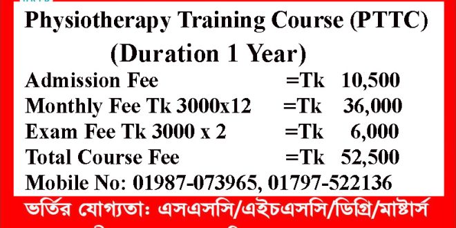
Physiotherapy Training Course (PTTC) 1 Year: Mobile No: 01969947171, 01987-073965, 01797-522136. This short course of Physiotherapy contains 10 subjects in 2 semesters. 1st Semester contains 5 subjects. The subjects are Human Anatomy & Physiology, Chemistry & Pharmacology, First Aid & Treatment, Hematology & Pathology, and the Study of OTC Drugs & Medicine.
2nd semester of this short physiotherapy course contains 5 subjects. The subjects are Orthopedic Anatomy & Physiology, Neuroanatomy & Physiology, Cardiovascular Anatomy & Physiology, Electro Physics, and the Study of TENS.
Course Fee Summary:
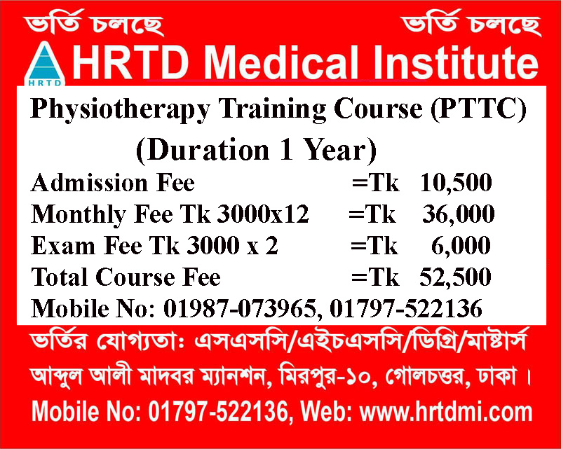
Course fee for 1 year Physiotherapy Training Course Tk 62,500/- including admission fee Tk 12,500/-, Monthly fee Tk 3500/- and exam fee Tk 3000×2 = 6000/-.
Location of Physiotherapy Training Course
Location of Physiotherapy Training Course. Mobile Phone Number 01969947171, 01797522136, 01987073965. HRTD Medical Institute, Section-6, Block-Kha, Road-1, Plot-11, Metro Rail Piller No. 249, Mirpur-10 Golchattar, Dhaka-1216. Mobile Phone No. 01797522136, 01987073965.
Objectives of Physiotherapy Course/ Physiotherapy Diploma
একজন ফিজিওথেরাপিষ্ট এর পক্ষে একটি ফিজিওথেরাপী চেম্বার বা ফিজিওথেরাপী সেন্টার বা ফিজিওথেরাপী ক্লিনিক ম্যানেজ করা সম্ভব হয় না বা ম্যানেজ করা উচিৎ না । কারন সেখানে থাকে অনেক মেশিনারী এবং অনেক কাজ । যেমন মেশিনারী ম্যানেজমেন্ট, মার্কেটিং ম্যানেজমেন্ট, একাউন্টস ম্যানেজমেন্ট, ফিনেনশিয়াল ম্যানেজমেন্ট, রোগীদের ডেটা ম্যানেজমেন্ট, রোগীদের কাউন্সিলিং ইত্যাদি ।
ফিজিওথেরাপি ৩ বছর/ ৪ বছরের ডিপ্লোমা কোর্স সফলভাবে সমাপ্ত করার পরে, শিক্ষার্থীরা সক্ষম হবে:

(১) ফিজিওথেরাপি সম্পর্কিত অনেক জ্ঞান ও দক্ষতা অর্জন করতে পারবে ।
(২) বিভিন্ন ফিজিওথেরাপি সরঞ্জাম এবং আনুষাঙ্গিক পরিচালনা এবং রক্ষণাবেক্ষণের জ্ঞান ও দক্ষতা অর্জন করতে পারবে ।
(৩) ফিজিওথেরাপি সম্পর্কিত প্রয়োজনীয় ডকুমেন্টেশন বজায় রাখার জ্ঞান ও দক্ষতা অর্জন করতে পারবে ।
(৪) ফিজিওথেরাপি যন্ত্রগুলি পরিচালনার বিপদ এবং তাদের প্রতিরক্ষামূলক জ্ঞান ও দক্ষতা অর্জন করতে পারবে ।
(৫) ফিজিওথেরাপিউটিক কার্যক্রম তদারকি জ্ঞান ও দক্ষতা অর্জন করতে পারবে ।

(৬) ফিজিওথেরাপিউটিক কার্যক্রমের ভবিষ্যত উন্নয়ন ও পরিকল্পনায় অবদান রাখাতে পারবে।
Admission Eligibility for Physiotherapy Training Courses in Dhaka
Admission Eligibility for Paramedical Courses. Mobile No. 01987-073965, 01797-522136. SSC Pass/ Equivalent/ HSC/ Degree/ Masters from arts, commerce, science, technical, etc.
Class Time for Physiotherapy Training Course
Weekly Class 3 hours. For Regular Students Friday 1 hour, Saturday 1 hour, and Monday 1 hour. For Job holders, Friday is 3 hours, or Monday is 3 hours. Morning Shift 9:00 am to 12:00 pm, and Evening Shift 3:00 pm to 6:00 pm.
ফিজিওথেরাপি ডিপ্লোমা বা ফিজিওথেরাপি কোর্স কেন করবেন?
ফিজিওথেরাপি ডিপ্লোমা বা ফিজিওথেরাপি কোর্স করে ফিজিওথেরাপি চ্যাম্বার, ফিজিওথেরাপি সেন্টার, ফিজিওথেরাপি ক্লিনিক, অথরা ফিজিওথেরাপি হাসপাতাল গড়ে তুলতে পারবেন। এটা নির্ভর করে আপনার অর্থনৈতিক অবস্থা এবং মেধার উপর । ফিজিওথেরাপি সেন্টারে চাকরি করতে পারবেন, বাসায় বাসায় অথবা হাসপাতালে হাসপাতালে গিয়ে থেরাপী দিতে পারবেন ।
ফিজিওথেরাপি ১ বছরের কোর্স এ কি কি শেখানো হবে?
১ বছরের ফিজিওথেরাপি সার্টিফিকেট কোর্সে সাধারণত প্রাথমিক চিকিৎসা, মানবদেহের গঠন, এবং বিভিন্ন শারীরিক সমস্যা দূর করার কৌশল শেখানো হয়। মূল বিষয়গুলোর মধ্যে রয়েছে মানবদেহের অস্থি ও মাংসপেশির পরিচিতি (Anatomy), থেরাপিউটিক এক্সারসাইজ বা ব্যায়াম, ইলেক্ট্রোথেরাপির ব্যবহার, এবং প্যারালাইসিস, বাত ও আঘাতজনিত ব্যথার পুনর্বাসন কৌশল।
কোর্সটির সাধারণ পাঠ্যসূচি ও কারিকুলাম নিচে দেওয়া হলো:
- বেসিক অ্যানাটমি এবং ফিজিওলজি: মানবদেহের হাড়, জয়েন্ট বা জোড়া এবং মাংসপেশির প্রাথমিক ধারণা।
- ইলেক্ট্রোথেরাপি (Electrotherapy): বিভিন্ন মেশিনের সাহায্যে ব্যথা কমানোর কৌশল, যেমন- Short Wave Diathermy, TENS।
- এক্সারসাইজ থেরাপি (Exercise Therapy): বিভিন্ন শারীরিক সমস্যা, স্পোর্টস ইনজুরি ও স্ট্রোকের পর নড়াচড়া উন্নত করার ব্যায়াম।
- ম্যানুয়াল থেরাপি এবং ম্যাসাজ: হাত দিয়ে শরীরের বিভিন্ন অংশের আড়ষ্টতা ও জয়েন্টের ব্যথা কমানোর পদ্ধতি।
- পুনর্বাসন (Rehabilitation): হাঁটাচলার জন্য সহায়ক সামগ্রী (যেমন- ওয়াকার, ক্রাচ) ব্যবহারের নিয়মকানুন এবং দৈনন্দিন কাজের ট্রেনিং।
- প্রাথমিক চিকিৎসা এবং রোগীর যত্ন (First Aid): জরুরি অবস্থায় করণীয় ও পেশেন্ট হ্যান্ডলিং।
Hostel Facilities in HRTD Medical Institute for Physiotherapy Training Course 1 Year
Hostal & Meal Facilities
The Institute has hostel facilities for the students. Students can take a bed in the hostel.
Hostel Fee Tk 3000/- Per Month
Meal Charges Tk 3000/- Per Month. ( Approximately )
হোস্টাল ও খাবার সুবিধা
ইনস্টিটিউটে শিক্ষার্থীদের জন্য হোস্টেল সুবিধা রয়েছে। ছাত্ররা হোস্টেলে বিছানা নিতে পারে।
হোস্টেল ফি 3000/- টাকা প্রতি মাসে,
খাবারের চার্জ 3000/- টাকা প্রতি মাসে।(প্রায়)
Teacher of Physiotherapy Training Course
- Dr. Md. Sakulur Rahman, MBBS, CCD (BIRDEM), Course Director
- Dr. Sanjana Binte Ahmed, BDS, MPH, Assistant Course Director
- Dr. Tisha, MBBS, PGT Gyne, Assistant Course Director
- Dr. Suhana, MBBS, PGT Medicine
- Dr. Danial Hoque, MBBS, C-Card
- Dr. Tisha, MBBS
- Dr. Afrin Jahan, MBBS, PGT Medicine
- Dr. Ananna, MBBS
- Dr. Lamia Afroze, MBBS
- Dr. Amena Afroze Anu, MBBS, PGT Gyne, Assistant Course Director
- Dr. Farhana Antara, MBBS,
- Dr. Nazmun Nahar Juthi, BDS, PGT
- Dr. Farhana Sharna, MBBS
- Dr. Bushra, MBBS
- Dr. Turzo, MBBS
- Dr. Kamrunnahar Keya, BDS, PGT (Dhaka Dental College)
- Dr. Shamima, MBBS, PGT Gyne
- Dr. Alamin, MBBS
- Dr. Benzir Belal, MBBS
- Dr. Disha, MBBS
- Dr. Mahinul Islam, MBBS
- Dr. Tisha, MBBS, PGT Medicine
- Dr. Anika, MBBS, PGT
- Dr. Jannatul Ferdous, MBBS, PGT Gyne
- Dr. Jannatul Aman, MBBS, PGT
- Dr. Rayhan, BPT
- Dr. Abu Hurayra, BPT
- Dr. Sharmin Ankhi, MBBS, PGT Medicine
- Md. Monir Hossain, B Pharm, M Pharm
- Md. Monirul Islam, B Pharm, M Pharm
- Md. Feroj Ahmed, BSc Pathology, PDT Medicine
Subjects for Physiotherapy Training Course
1st Semester Subject:
Human Anatomy & Physiology
Chemistry & Pharmacology
First Aid & Treatment
Hematology & Pathology
Orthopedic Anatomy & Physiology
2nd Semester Subject:
Neuro Anatomy & Physiology
Orthopedic Disease & Complication
Therapeutic Exercise
Electro Physics
Electrotherapy
Human Anatomy & Physiology for Physiotherapy Training Course
Human Anatomy & Physiology is an important subject in physiotherapy courses. The study of Body Structure and its functions is Anatomy and Physiology. Here we discuss the systems of the Human Body and its Organs, Tissues, and Cells. The systems of the Human Body are the Nervous System, Digestive System, Respiratory System, Circulatory System or Cardiovascular System, Skeletal System, Muscular System, Endocrine System, Immune System, Lymphatic System, Integumentary System or Covering System, and Urinary System.
শারীরিক গঠন এবং এর কার্যাবলীর অধ্যয়ন হল অ্যানাটমি এবং ফিজিওলজি। এখানে আমরা মানবদেহের সিস্টেম এবং এর অঙ্গ, টিস্যু এবং কোষ নিয়ে আলোচনা করি। মানবদেহের সিস্টেমগুলি হ’ল হজম সিস্টেম, শ্বাসযন্ত্র, কার্ডিওভাসকুলার সিস্টেম, কঙ্কাল সিস্টেম, পেশীতন্ত্র, স্নায়ুতন্ত্র, এন্ডোক্রাইন সিস্টেম, ইমিউন সিস্টেম, লিম্ফ্যাটিক সিস্টেম, ইন্টিগুমেন্টারি সিস্টেম এবং ইউরিনারি সিস্টেম।
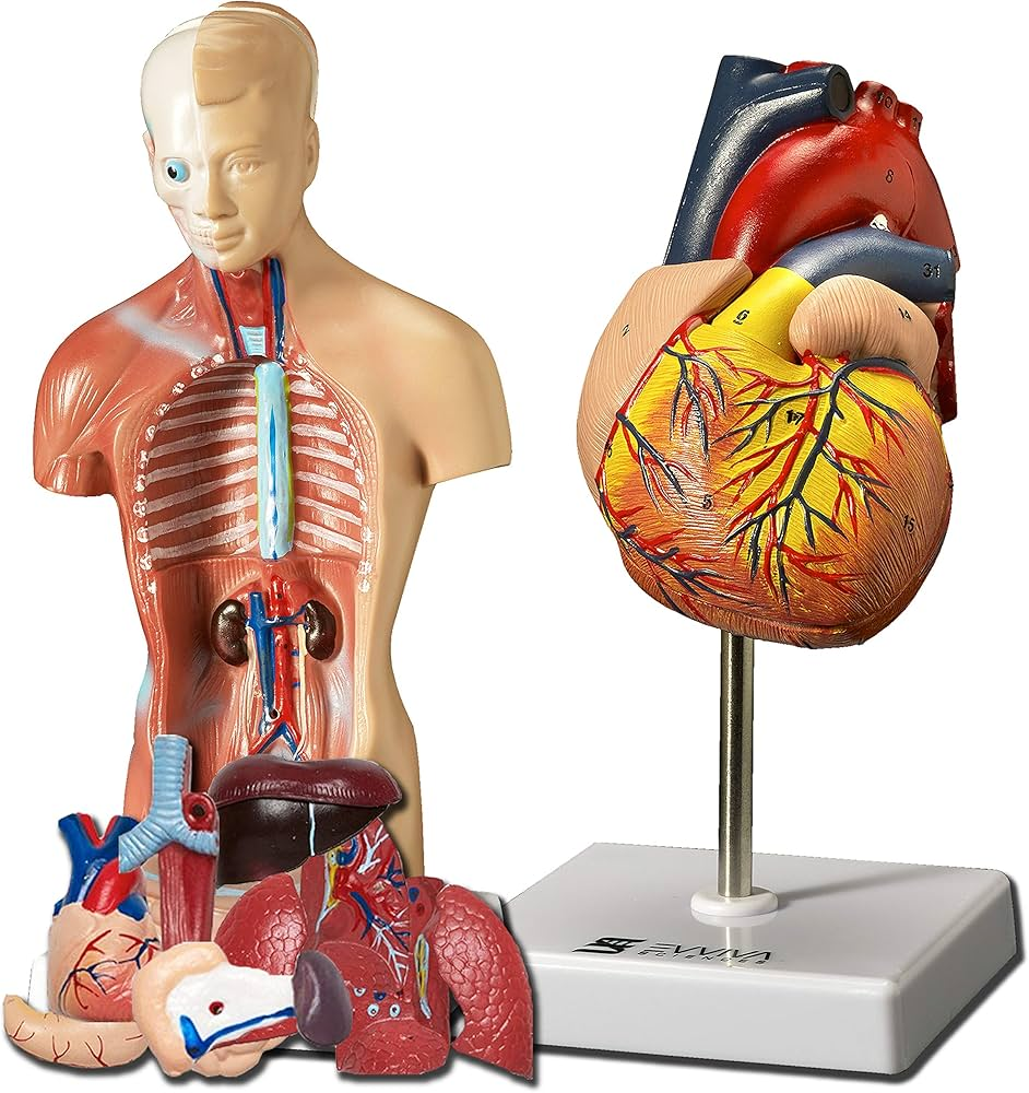
Anatomy is the scientific study of the structural organization and physical relationships of living things and their parts. Derived from the Greek word anatome, meaning “to cut up” or “dissection,” it describes where body structures are located and what they are made of.
Physiology is the scientific study of the functions and mechanisms of living systems. While anatomy focuses on structure, physiology focuses on how those structures—from molecules to organ systems—work together to support life.
The human body is organized into 11 major organ systems that work together to maintain homeostasis.
1. Integumentary System
- Primary Function: Forms a protective physical barrier, regulates body temperature, and detects sensory stimuli.
- Key Components: Skin (epidermis, dermis, hypodermis), hair, nails, and exocrine glands (sweat, oil).
- Physiology: Synthesizes Vitamin D from sunlight and eliminates waste through perspiration.
2. Skeletal System
- Primary Function: Provides structural support, protects vital organs, and facilitates movement.
- Key Components: Bones (206 in adults), cartilage, ligaments, and joints.
- Physiology: Red bone marrow produces blood cells (hematopoiesis), while the bone matrix stores essential minerals like calcium and phosphate.
3. Muscular System
- Primary Function: Facilitates voluntary and involuntary movement and generates body heat.
- Key Components: Skeletal muscles, smooth muscles (in organs), and cardiac muscle (heart).
- Physiology: Muscles pull on bones via tendons to create leverage for locomotion.
4. Nervous System
- Primary Function: Coordinates rapid responses to internal and external stimuli through electrical signaling.
- Key Components: Brain, spinal cord, nerves, and sensory organs (eyes, ears).
- Physiology: Divided into the Central Nervous System (CNS) for processing and the Peripheral Nervous System (PNS) for transmission.
5. Endocrine System
- Primary Function: Regulates long-term processes like growth, metabolism, and reproduction through hormones.
- Key Components: Glands such as the pituitary, thyroid, adrenal, pancreas, and gonads.
- Physiology: Hormones are secreted directly into the bloodstream to target distant organs.
6. Cardiovascular (Circulatory) System
- Primary Function: Transports oxygen, nutrients, hormones, and waste throughout the body.
- Key Components: Heart, blood vessels (arteries, veins, capillaries), and blood.
- Physiology: Operates in two main circuits—pulmonary (lungs) and systemic (rest of the body).
7. Lymphatic and Immune System
- Primary Function: Drains excess tissue fluid (lymph) back to the blood and defends against pathogens.
- Key Components: Lymph nodes, spleen, thymus, tonsils, and lymphatic vessels.
- Physiology: Lymphocytes (white blood cells) sample lymph for invaders to mount immune responses.
8. Respiratory System
- Primary Function: Facilitates gas exchange, bringing in oxygen and expelling carbon dioxide.
- Key Components: Lungs, trachea, pharynx, larynx, and diaphragm.
- Physiology: Gas exchange occurs across the thin membranes of the alveoli in the lungs.
9. Digestive System
- Primary Function: Breaks down food mechanically and chemically for nutrient absorption.
- Key Components: Mouth, esophagus, stomach, small and large intestines, liver, and pancreas.
- Physiology: Chyme (processed food) moves through the small intestine where villi absorb nutrients into the blood.
10. Urinary (Excretory) System
- Primary Function: Filters blood to eliminate metabolic waste and regulates fluid and electrolyte balance.
- Key Components: Kidneys, ureters, urinary bladder, and urethra.
- Physiology: Nephrons in the kidneys purify blood and produce urine for excretion.
11. Reproductive System
- Primary Function: Produces gametes (sperm/eggs) and sex hormones for the production of offspring.
- Key Components: Testes, ovaries, uterus, and associated ducts/glands.
- Physiology: Regulated by the endocrine system to manage cycles like menstruation or sperm production.
Pharmacology for Physiotherapy Training Course
The study of Drugs and Medicine is called Pharmacology. Drugs are chemicals that can react with cells, tissues, and organs of the body. Here we discuss group-wise drugs and their medicines. Common Groups of Drugs are Pain Killer Drugs, Anti Ulcer Drugs, Anti Vomiting Drugs, Anti Viral Drugs, Laxative Drugs, Motility Drugs and Antimotility Drugs, Bronchodilator Drugs, Antibiotic Drugs, Anti Fungal Drugs, Anti Thrombotic Drugs, Anti Protozoal Drugs, Anthelmintic Drugs, Anti Hypertensive Drugs, Beta Blocker Drugs, Calcium Channel Blocker Drugs, ACE Inhibitor Drugs, Hemostatic Drugs, Analgesic Drugs, Antipyretic Drugs, Steroid Drugs, NSAID Drugs, CNS Drugs, Neuropathic Pain Keller Drugs, Spasmodic Drugs, etc.
ওষুধ ও ওষুধের অধ্যয়নকে ফার্মাকোলজি বলা হয়। এখানে আমরা ফার্মাকোলজি-১ এ গ্রুপভিত্তিক ওষুধ ও তাদের ওষুধ নিয়ে আলোচনা করেছি। ওষুধের সাধারণ গ্রুপগুলি হল পেইন কিলার ড্রাগস, অ্যান্টি আলসার ড্রাগস, অ্যান্টি-ভোমিটিং ড্রাগস, ল্যাক্সেটিভ ড্রাগস, মোটিলিটি ড্রাগস, অ্যান্টিমোটিলিটি ড্রাগস, ব্রঙ্কোডাইলেটর ড্রাগস, অ্যান্টিবায়োটিক ড্রাগস, অ্যান্টি ফাংগাল ড্রাগস, অ্যান্টি প্রোটোজোয়াল ড্রাগস, অ্যান্টি ভাইরাল ড্রাগস, অ্যান্টি-ভাইরাল ড্রাগস, অ্যান্টি-অ্যান্টিভেনস ড্রাগস। , বিটা ব্লকার ওষুধ, ক্যালসিয়াম চ্যানেল ব্লকার ড্রাগস, এসিই ইনহিবিটর ড্রাগস, হেমোস্ট্যাটিক ড্রাগস, অ্যানালজেসিক ড্রাগস, অ্যান্টিপাইরেটিক ড্রাগস, অ্যান্টি থ্রম্বোটিক ড্রাগস ইত্যাদি।
Pharmacology for physiotherapy is a specialized field focused on how medications interact with physical rehabilitation. In 2025, courses increasingly emphasize clinical reasoning to manage patients on multiple medications (polypharmacy) and optimize treatment timing based on drug effects.
1. Core Pharmacological Concepts
- Pharmacokinetics (ADME): The study of what the body does to a drug, including Absorption, Distribution, Metabolism, and Excretion. For physiotherapists, this includes how exercise affects these processes (e.g., increased blood flow altering drug absorption).
- Pharmacodynamics: The study of what a drug does to the body, focusing on drug-receptor interactions and therapeutic versus toxic effects.
- Therapeutic Index: A measure of a drug’s safety; drugs with a narrow index require close monitoring for adverse reactions during physical activity.
2. Major Drug Classes in Physiotherapy
Physiotherapy courses typically cover medications that directly impact movement, pain, and exercise tolerance:
- Analgesics & Anti-inflammatories: NSAIDs (ibuprofen), acetaminophen, and opioids used for pain management. These can mask symptoms, potentially leading to overexertion during therapy.
- Cardiovascular Drugs: Beta-blockers, ACE inhibitors, and diuretics. These affect heart rate and blood pressure, often requiring intensity adjustments during aerobic training.
- Neurological & Psychotropic Drugs: SSRIs, muscle relaxants, and anti-Parkinsonian agents. These can cause side effects like dizziness, drowsiness, or impaired balance, significantly increasing fall risks.
- Endocrine Drugs: Metformin and insulin. Physiotherapists must monitor for hypoglycemia (low blood sugar) triggered by exercise.
3. Clinical Implications for Practice
Modern 2025 curricula focus on integrating drug knowledge into patient safety and treatment planning:
- Treatment Timing: Scheduling sessions when analgesics are at peak effect for better performance, or avoiding times when sedatives are strongest to prevent falls.
- Red Flag Screening: Identifying medication-related “red flags” such as GI bleeding (from NSAIDs), orthostatic hypotension (from antihypertensives), or muscle myopathy (from statins).
- Interprofessional Communication: Using structured formats like SBAR to report suspected adverse drug reactions to prescribing physicians.
- Patient Education: Advising patients on medication adherence and the functional impact of their prescriptions on rehabilitation goals.
First Aid for Physiotherapy Training Course
First Aid is an important subject for all courses. It is very important for the Physiotherapy Training Course. Here we discuss Shock, Classifications of Shock, Causes of Shock, Stages of Shock, Clinical Features of Shock, Hypovolemic Shock, Cardiogenic Shock, Neurogenic Shock, Traumatic Shock, Burn Shock, Electric Shock, Psychogenic Shock, Anaphylactic Shock, First Aid of Shock, Management of Shock, First Aid of Cut, First of Snake Bite, First Aid of Accidental Injury, etc.
ফার্স্ট এইড মেডিসিন এবং সার্জারি কোর্সের ডিপ্লোমা, আরএমপি কোর্স, এলএমএএফ কোর্স, প্যারামেডিক্যাল কোর্স, ডিএমএ কোর্স, ডিএমএস কোর্স, নার্সিং কোর্স, ডেন্টাল কোর্স, প্যাথলজি কোর্স, ফিজিওথেরাপি কোর্স, কেয়ারগিভার কোর্স, ইত্যাদির জন্য একটি গুরুত্বপূর্ণ বিষয়। শক, শ্রেণিবিন্যাস শক, শকের কারণ, পর্যায় নিয়ে আলোচনা করুন শক, শকের ক্লিনিকাল বৈশিষ্ট্য, হাইপোভোলেমিক শক, কার্ডিওজেনিক শক, নিউরোজেনিক শক, ট্রমাটিক শক, বার্ন শক, বৈদ্যুতিক শক, সাইকোজেনিক শক, অ্যানাফিল্যাকটিক শক, শকের প্রাথমিক চিকিৎসা, কাটার প্রাথমিক চিকিৎসা, সাপের কামড়ের প্রাথমিক চিকিৎসা, প্রাথমিক চিকিৎসা দুর্ঘটনাজনিত আঘাত, ইত্যাদি
First Aid training for physiotherapy students and professionals focuses on integrating emergency life support with musculoskeletal expertise. Training typically covers both standard emergency response and clinical risks specific to the physiotherapy environment.
1. Core Training Components (2025 Standards)
Standard physiotherapy curricula in 2025 include First Aid as a foundational subject. Key learning points include:
- Emergency Life Support: Management of an unconscious casualty, recovery positioning, and high-quality CPR for adults, children, and infants.
- Cardiac Care: Recognition and management of heart attacks, angina, and the immediate use of an Automated External Defibrillator (AED).
- Acute Trauma & Injuries: Treatment for major/minor bleeding, burns, head and spinal injuries (concussion/compression), and bone fractures.
- Soft Tissue Management: Immediate care for sprains, strains, and dislocations using protocols like PRICE (Protection, Rest, Ice, Compression, Elevation).
- Medical Emergencies: Recognizing and treating anaphylaxis (allergic reactions), asthma attacks, strokes, seizures, and signs of sepsis or meningitis.
2. Specialized Requirements for Physiotherapists
- Mandatory Competencies: Physiotherapists working under Sports Physiotherapy Standards must maintain up-to-date competencies in basic life support and acute trauma management.
- Renewals & CPD: First Aid certificates are typically valid for 3 years, though Basic Life Support (BLS) or CPR-specific refreshers are often required annually in clinical settings.
- 2025 Certification Update: Starting in 2025, some regulatory bodies now require BLS CPR (specifically) for professional renewals, which may also qualify for increased Professional Development Credits (PDCs).
3. Training Formats and Options
Physiotherapy-specific courses are often tailored to the clinical environment:
- 3-Hour Refresher: Focused on CPR, AED use, heart attacks, and choking.
- Emergency First Aid at Work (EFAW): A 1-day (or online level 3) course for lower-risk clinical environments.
- Advanced First Aid: Comprehensive 70-80+ hour programs for professionals in high-risk or industrial settings, covering advanced patient assessment and transportation.
- Blended Learning: Many 2025 providers offer online theory modules followed by practical, hands-on skill sessions to fit around clinical schedules.
Study of OTC Drugs for Physiotherapy Training Course
OTC is the short form of over-the-counter. That is OTC Drugs are over-the-counter drugs. The study of OTC Drugs is important for Physiotherapy Courses. These Drugs can be sold or purchased without any prescription from Registered MBBS Doctors. These Drugs are Emergency and Safe for the patients. The study of OTC Drugs improves the quality of practice. Some OTC Drugs are Paracetamol, Albendazole, Ascorbic Acid, Calcium, Multivitamins, Vitamin B Complex, Omeprazole, Oral Rehydration Salt, Salbutamol, Mebendazole, Neomycin, Gentamycin, Bacitracin, etc.
ওটিসি ওষুধগুলি সমস্ত মেডিকেল কোর্স,ডিপ্লোমা মেডিকেল কোর্স, এলএমএএফ কোর্স এবং আরএমপি কোর্সের জন্য গুরুত্বপূর্ণ। ডিপ্লোমা ইন ফিজিওথেরাপির জন্যও এটি গুরুত্বপূর্ণ। এই OTC ওষুধগুলি নিবন্ধিত এমবিবিএস ডাক্তারদের কোনও প্রেসক্রিপশন ছাড়াই বিক্রি বা কেনা যায়। এই ওষুধগুলি জরুরী এবং রোগীদের জন্য নিরাপদ। ওটিসি ওষুধের অধ্যয়ন অনুশীলনের মান উন্নত করে। কিছু ওটিসি ওষুধ হল অ্যালবেনডাজল, অ্যাসকরবিক অ্যাসিড, ক্যালসিয়াম, মাল্টিভিটামিন, ভিটামিন বি কমপ্লেক্স, ওমেপ্রাজল, ওরাল রিহাইড্রেশন সল্ট, সালবুটামল ইত্যাদি।
Hematology and Pathology for Physiotherapy Training Course
Hematology and Pathology are important subjects in medical science. The study of Blood is called Hematology and the Study of Pathos is called Pathology. In Hematology and Pathology, we discuss blood cells, their morphology and functions, Blood Diseases, Common Pathos and their pathogenesis, Atrophy, Hypertrophy, Metaplasia, Gangrene, Pathological Tests like TC, DC, ESR, Hemoglobin Percentage, etc.
রক্ত ও রক্তের রোগের অধ্যয়নকে বলা হয় হেমাটোলজি এবং স্টাডি অফ প্যাথোস এবং রোগ সৃষ্টি ও নির্ণয়ের প্রক্রিয়াকে প্যাথলজি বলা হয়। হেমাটোলজি এবং প্যাথলজিতে, আমরা রক্তের কোষ, তাদের গঠন এবং কার্যকারিতা, রক্তের রোগ, সাধারণ প্যাথোস এবং তাদের প্যাথোজেনেসিস, অ্যাট্রোফি, হাইপারট্রফি, মেটাপ্লাসিয়া, গ্যাংগ্রিন, প্যাথলজিকাল টেস্ট যেমন TC, DC, ESR, হিমোগ্লোবিন শতাংশ ইত্যাদি নিয়ে আলোচনা করি।
Core Hematology Topics in Physiotherapy
The curriculum is designed to help you safely manage patients who have blood disorders or compromised circulatory systems. Key areas of study include:
- Blood Components and Physiology: Understanding the formation of blood cells (hematopoiesis), plasma, red blood cells (RBCs), white blood cells (WBCs), and platelets.
- Complete Blood Count (CBC) Interpretation: Learning to read and evaluate lab values, which is critical for determining if a patient is medically stable enough for exercise or manual therapy.
- Anemias: Studying different types of anemia (e.g., iron deficiency, sickle cell) and understanding how they cause fatigue and reduced exercise tolerance.
- Hemostasis and Coagulation: Learning about blood clotting, clotting factors, and bleeding disorders. This is vital when treating patients on blood thinners to avoid excessive bruising or bleeding during deep tissue therapies. [
- Malignancies: Identifying blood-related cancers like leukemia and lymphomas, and understanding their implications for safe physical therapy and rehabilitation.
Treatment/Practice of Medicine for Physiotherapy Training Course
The study of Disease and Treatment is called the Practice of Medicine. This subject is important for a Diploma Medical Practitioner, Diploma Medical Assistant, and Rural Medical Practitioner. This subject discusses some common diseases. The discussion points for the Practice of Medicine are the Definition of Disease, Causes of Disease, Clinical Features of Disease ( Symptoms and Signs), Investigation of Disease, Treatment of Disease, Complication of Disease, and Advice for the Patients. To understand the diseases, the movement of doctors, and their prescriptions, this subject should be studied by physiotherapy students.
রোগ ও চিকিৎসার অধ্যয়নকে বলা হয় মেডিসিনের অনুশীলন। এই বিষয় একজন ডিপ্লোমা মেডিকেল প্র্যাকটিশনার, ডিপ্লোমা মেডিকেল অ্যাসিস্ট্যান্ট এবং গ্রামীণ চিকিৎসা অনুশীলনকারীর জন্য গুরুত্বপূর্ণ। এই বিষয়ে কিছু সাধারণ রোগ নিয়ে আলোচনা করা হয়েছে। মেডিসিন অনুশীলনের আলোচনার বিষয়গুলি হল রোগের সংজ্ঞা, রোগের কারণ, রোগের ক্লিনিক্যাল বৈশিষ্ট্য (লক্ষণ এবং লক্ষণ), রোগের তদন্ত, রোগের চিকিৎসা, রোগের জটিলতা এবং রোগীদের জন্য পরামর্শ। রোগগুলি, ডাক্তারদের গতিবিধি এবং তাদের প্রেসক্রিপশন বোঝার জন্য, এই বিষয়টি ফিজিওথেরাপি শিক্ষার্থীদের দ্বারা অধ্যয়ন করা উচিত।
Therapeutic Exercise-1 for Physiotherapy Training Course
Exercise Therapies are discussed in Therapeutic Exercise. After completing Theory Classes, Practical Classes are done. Topics for Therapeutic Exercise-1 are Lever, Types of Lever, 1st Degree Lever, Pulley, Types of Pulley, Movable Pulley, Energy, Types of Energy, Kinetic Energy, Axis, Types of Axis, Plane, Types of Plane, Names of Shoulder Movement, Names of Hip Movement, Names of Wrist Movement, MCP Movement, Range of Motion of Elbow Joint, Muscle action, Types of Muscle action, Muscle work, Types of Muscle work, Types of Fundamental Position, Pelvic tilt, Types o Pelvic tilt, Relax Passive Movement, Indication of Relax Passive Movement, Contraindication of Relax Passive Movement.
ব্যায়াম থেরাপি থেরাপিউটিক ব্যায়াম আলোচনা করা হয়. থিওরি ক্লাস শেষ করার পর ব্যবহারিক ক্লাস করা হয়। থেরাপিউটিক এক্সারসাইজ-১-এর বিষয়গুলো হলো লিভার, লিভারের ধরন, ১ম ডিগ্রি লিভার, পুলি, পুলির প্রকার, চলনশীল পুলি, শক্তি, শক্তির প্রকার, গতিশক্তি, অক্ষ, অক্ষের প্রকার, সমতল, সমতলের ধরন, নাপুল। আন্দোলন, নিতম্ব আন্দোলনের নাম, কব্জির নড়াচড়ার নাম, MCP মুভমেন্ট, কনুই জয়েন্টের গতির পরিসর, পেশীর ক্রিয়া, পেশীর ক্রিয়ার ধরন, পেশীর কাজ, পেশীর কাজের ধরন, মৌলিক অবস্থানের ধরন, শ্রোণী কাত, পেলভিক কাত, শিথিলকরণ, নিষ্ক্রিয় গতির ধরন প্যাসিভ আন্দোলন শিথিল করুন, প্যাসিভ মুভমেন্ট শিথিল করার বিরোধীতা,
Types of skeletal muscle fibers, Clinical Examination of the Motor Nervous System, Clinical Examination of the Sensory Nervous System, Clinical Examination of Cranial Nerves, Clinical Examination of the Cardiovascular System, Measurement of the Pelvic Angle of Inclination, Factors affecting the Joint Range of Motion, Starting and Derived Positions, Suspension therapy, Advantage of Suspension therapy, Names of Suspension Instrument, Types of Suspension Therapy, etc.
কঙ্কালের পেশী তন্তুর ধরন, মোটর স্নায়ুতন্ত্রের ক্লিনিকাল পরীক্ষা, সংবেদনশীল স্নায়ুতন্ত্রের ক্লিনিকাল পরীক্ষা, ক্রানিয়াল স্নায়ুর ক্লিনিকাল পরীক্ষা, কার্ডিওভাসকুলার সিস্টেমের ক্লিনিক্যাল পরীক্ষা, পেলভিক প্রবণতার কোণ পরিমাপ, জোড়ের সীমাকে প্রভাবিত করার কারণগুলি , শুরু এবং প্রাপ্ত অবস্থান, সাসপেনশন থেরাপি, সাসপেনশন থেরাপির সুবিধা, সাসপেনশন যন্ত্রের নাম, সাসপেনশন থেরাপির প্রকারভেদ ইত্যাদি।
Electro Therapy for Physiotherapy Training Course
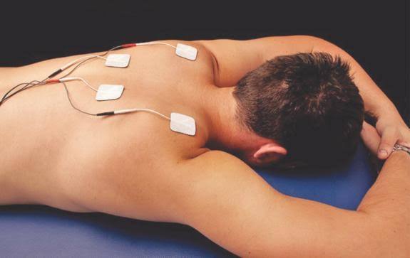
Electrotherapy uses electrical currents for medical treatment, primarily to relieve pain, reduce inflammation, and stimulate muscles, commonly seen in physiotherapy with devices like TENS or NMES, but also extends to psychiatric uses like Electroconvulsive Therapy (ECT) for severe mood disorders, and even cosmetic treatments for skin. It works by applying electrical energy to tissues, affecting nerve signals or muscle contractions for therapeutic effects, often complementing other active therapies for better results.
Common Types & Uses:
- Transcutaneous Electrical Nerve Stimulation (TENS): Mild currents through skin pads to block pain signals (gate control theory).
- Neuromuscular Electrical Stimulation (NMES): Stronger currents to contract muscles, preventing atrophy, retraining muscles after injury, or improving strength.
- Interferential Current (IFC): Uses two currents for deeper pain relief and increased circulation.
- Electroconvulsive Therapy (ECT): Controlled electric seizure for severe depression, bipolar disorder, or catatonia, when other treatments fail.
- Galvanic Current (Cosmetic): Uses direct current for skin cleansing (desincrustation) and nourishing (iontophoresis) to improve skin conditions like aging or acne.
How it Works (General):
- Pain Relief: Interrupts pain signals or triggers endorphin release.
- Muscle Rehab: Stimulates nerve & muscle fibers, improving tone, strength, or re-education.
- Healing: Can accelerate healing and reduce swelling by improving local circulation.
Key Considerations:
- Not a Replacement: Best when combined with exercise and manual therapy, not as a standalone solution.
- Skilled Application: Effectiveness depends on the therapist choosing the right type, intensity, and duration for the individual.
Cardiovascular Anatomy & Physiology for Physiotherapy Training Course
Cardiovascular Anatomy is a branch of Anatomy, and Cardiovascular Physiology is a branch of Physiology. These two subjects are related to cardiology. Cardiovascular Anatomy and Physiology are being studied in a single subject for Diploma Medical Courses, Paramedical Courses, and All Short Medical Courses.
কার্ডিওভাসকুলার অ্যানাটমি হল অ্যানাটমির একটি শাখা এবং কার্ডিওভাসকুলার ফিজিওলজি হল ফিজিওলজির একটি শাখা। এই দুটি বিষয় কার্ডিওলজি সম্পর্কিত। কার্ডিওভাসকুলার অ্যানাটমি এবং ফিজিওলজি ডিপ্লোমা মেডিকেল কোর্স, প্যারামেডিক্যাল কোর্স এবং সমস্ত শর্ট মেডিকেল কোর্সের জন্য একটি একক বিষয়ে অধ্যয়ন করা হচ্ছে।
We discuss here the Anatomy of the Heart, Cardiac Chambers, Cardiac Valves, Cardiac Wall, Cardiac Septum, Right Heart, Left Heat, Function of Right Heat, Functions of Left Heart, Aorta, Venecava, Artery, Vein, Capillary, Pulmonary Blood Circulation, Cerebral Blood Circulation, Renal Blood Circulation, Hepatic Blood Circulation, Portal Vein and Portal Circulation, Heart Beat, Pulse, Pulse Rate, Tachycardia, Bradycardia, Blood Pressure, Normal Blood Pressure, Hypertension, Hypotension, Stroke Volume, Cardiac Output, Heart Failure, etc. This Subject is the most essential for the Diploma in Medicine and Diploma in Surgery Course.
আমরা এখানে হার্টের অ্যানাটমি, কার্ডিয়াক চেম্বার, কার্ডিয়াক ভালভ, কার্ডিয়াক ওয়াল, কার্ডিয়াক সেপ্টাম, ডান হার্ট, বাম তাপ, ডান তাপের ফাংশন, বাম হার্টের কাজ, অ্যাওর্টা, ভেনেকাভা, ধমনী, শিরা, কৈশিক, পালমোনারি রক্ত সঞ্চালন নিয়ে আলোচনা করি। , সেরিব্রাল ব্লাড সার্কুলেশন, রেনাল ব্লাড সার্কুলেশন, হেপাটিক ব্লাড সঞ্চালন, পোর্টাল ভেইন এবং পোর্টাল সঞ্চালন, হার্ট বিট, পালস, পালস রেট, টাকাইকার্ডিয়া, ব্র্যাডিকার্ডিয়া, রক্তচাপ, স্বাভাবিক রক্তচাপ, উচ্চ রক্তচাপ, হাইপোটেনশন, স্ট্রোক ভলিউম, কার্ডিয়াক আউটপুট, হার্ট ফেইলিউর ইত্যাদি। ডিপ্লোমা ইন মেডিসিন এবং ডিপ্লোমা ইন সার্জারি কোর্স।
Orthopedic Anatomy & Physiology for Physiotherapy Training Course
Orthopedic Anatomy & Physiology describe the anatomy of bone, physiology of bone, anatomy of skeletal muscle, physiology of skeletal muscle, anatomy of tendon, physiology of tendon, anatomy of ligament physiology of ligament, anatomy of cartilage, physiology of cartilage, anatomy of joints, physiology of joints, etc.
অর্থোপেডিক অ্যানাটমি এবং ফিজিওলজি হাড়ের অ্যানাটমি, হাড়ের শারীরবৃত্তি, কঙ্কালের পেশীর শারীরবৃত্তি, কঙ্কালের পেশীর শারীরবৃত্তি, টেন্ডনের শারীরবৃত্তি, টেন্ডনের শারীরবৃত্তি, লিগামেন্টের অ্যানাটমি অফ লিগামেন্ট ফিজিওলজি, লিগামেন্টের অ্যানাটমি, কারটিলেজের শারীরবৃত্তি, কার্টিলেজের শারীরবৃত্তবিদ্যা, কার্টিলেজের শারীরবৃত্তির বর্ণনা দেয়। জয়েন্ট, ইত্যাদি
Orthopedic Disease & Complications for Physiotherapy Training Course
Orthopedic disease refers to any disorder, injury, or condition affecting the musculoskeletal system, which includes bones, joints, muscles, ligaments, tendons, and cartilage. These diseases may cause pain, deformity, limitation of movement, and functional disability.
👉 Physiotherapy focus:
Orthopedic diseases are treated with exercise therapy, electrotherapy, manual therapy, and rehabilitation techniques to restore movement, strength, and daily function.
1. Fracture
Explanation:
A fracture is a break or crack in a bone caused by trauma, fall, or accident. It leads to pain, swelling, and inability to move the affected part.
Types
- Simple (Closed)
- Compound (Open)
- Comminuted
- Greenstick
- Stress fracture
Medical Treatment
- Immobilization (cast, splint)
- Traction
- Surgery (ORIF – plates, screws, nails)
Physiotherapy Treatment
- Pain & edema control (ICE, TENS)
- ROM exercises
- Isometric → isotonic strengthening
- Gait training
- Functional rehabilitation
2. Osteoarthritis
Explanation:
Osteoarthritis is a degenerative joint disease where the cartilage between joints wears out, causing pain, stiffness, and reduced movement.
Common Joints
- Knee
- Hip
- Spine
- Hand
Medical Treatment
- NSAIDs
- Analgesics
- Weight reduction
- Joint replacement (severe cases)
Physiotherapy Treatment
- Hot therapy
- Quadriceps strengthening
- ROM exercises
- Low-impact aerobic exercise
- Joint protection education
3. Rheumatoid Arthritis
Explanation:
Rheumatoid arthritis is a chronic autoimmune disease that causes inflammation of joints, leading to pain, swelling, and deformity.
Symptoms
- Morning stiffness
- Symmetrical joint pain
- Swelling & deformity
Medical Treatment
- DMARDs
- NSAIDs
- Steroids
Physiotherapy Treatment
- Pain relief modalities
- Gentle ROM
- Muscle strengthening
- Splinting
- Energy conservation techniques
4. Low Back Pain
Explanation:
Low back pain is discomfort in the lower spine region, often caused by muscle strain, poor posture, or disc problems.
Causes
- Muscle strain
- Disc prolapse
- Poor posture
Medical Treatment
- Analgesics
- Muscle relaxants
- Rest (short term)
Physiotherapy Treatment
- Heat therapy
- Core strengthening
- McKenzie exercises
- Posture correction
- Ergonomic advice
5. Cervical Spondylosis
Explanation:
Cervical spondylosis is degeneration of the cervical spine that causes neck pain, stiffness, and sometimes arm pain.
Symptoms
- Neck pain
- Stiffness
- Radiating arm pain
Medical Treatment
- Pain medication
- Cervical collar
Physiotherapy Treatment
- Cervical traction
- ROM exercises
- Isometric neck exercises
- Postural correction
6. Disc Prolapse (Slipped Disc)
Explanation:
Disc prolapse occurs when the intervertebral disc bulges out and presses on nerves, causing radiating pain and numbness.
Symptoms
- Radiating pain
- Numbness
- Muscle weakness
Medical Treatment
- Bed rest
- NSAIDs
- Surgery (severe cases)
Physiotherapy Treatment
- Traction
- McKenzie exercises
- Core strengthening
- Neural mobilization
7. Osteoporosis
Explanation:
Osteoporosis is a condition where bone density decreases, making bones weak and prone to fractures.
Medical Treatment
- Calcium & Vitamin D
- Bisphosphonates
Physiotherapy Treatment
- Weight-bearing exercises
- Balance training
- Fall prevention
- Postural exercises
8. Frozen Shoulder
Explanation:
Frozen shoulder is a condition characterized by pain and stiffness of the shoulder joint with restricted movement.
Medical Treatment
- Analgesics
- Steroid injection
Physiotherapy Treatment
- Hot therapy
- Pendular exercises
- Stretching
- Joint mobilization
9. Ligament Injury
Explanation:
Ligament injury occurs due to overstretching or tearing of ligaments, commonly seen in sports injuries like ankle sprain or ACL tear.
Medical Treatment
- RICE
- Bracing
- Surgery (severe)
Physiotherapy Treatment
- Strengthening
- Proprioceptive training
- Balance exercises
- Sport-specific rehab
10. Tendinitis
Explanation:
Tendinitis is inflammation of a tendon caused by overuse, leading to pain during movement.
11. Scoliosis
Explanation:
Scoliosis is an abnormal sideways curvature of the spine, which can affect posture and breathing.
12. Kyphosis
Explanation:
Kyphosis is excessive forward curvature of the thoracic spine, resulting in a hunched back.
13. Lordosis
Explanation:
Lordosis is excessive inward curvature of the lumbar spine, often associated with poor posture.
14. Gout
Explanation:
Gout is a type of arthritis caused by uric acid crystal deposition in joints, resulting in sudden severe pain and swelling.
15. Postural Deformity
Explanation:
Postural deformity occurs due to poor posture habits, causing abnormal alignment of the body and musculoskeletal pain.
Practicle Class for Physiotherapy Training Course 1 Year in Bangladesh
Manual /Exercise Therapy :
Surface Anatomy & basic assessment , AROM, PROM, AAROM , Muscle Power testing , Basic Stretching and positioning
Electro therapy
Electroduction to electro therapy instruments. Safety, Contraindication, TENS : Applied & Parameter
Manual Practicle Class in Surface Anatomy & basic assessment
Surface anatomy is the study of external bodily features used by physiotherapists to locate underlying muscles, bones, and joints. Mastery of palpation and visual landmarks enables clinicians to accurately perform physical examinations, identify painful structures, and safely guide therapeutic interventions like joint mobilizations and massage.
1. Palpation Techniques
Palpation is the cornerstone of a physiotherapy assessment. Use the pads of your index, middle, and ring fingers rather than your fingertips.
- Tissue Differentiation: Bones are firm and rigid. Tendons feel like tight, taut cords. Ligaments are dense, tough bands. Muscle bellies are softer and yield to pressure.
- Guidelines: Ensure your peer/model is in a relaxed position. Visualize the underlying structure in your mind before applying pressure, and start with light touch to assess skin temperature and texture before pressing deeply to locate bone or tissue.
2. Key Anatomical Landmarks
Familiarity with bony and soft-tissue landmarks allows you to identify spinal segments and joint lines quickly:
Spine
- C7 (Vertebra Prominens): Located at the base of the neck. It is the most prominent spinous process when the neck is flexed.
- T3 Spinous Process: Aligned with the medial root of the spine of the scapula.
- T7 Spinous Process: Aligned with the inferior angle of the scapula.
- L4 Spinous Process: Found by drawing a horizontal line across the highest points of the iliac crests (supracristal plane). This is the key landmark for lumbar spine levels.
- Posterior Superior Iliac Spine (PSIS): Visible as dimples of Venus; directly over the S2 sacral segment.
Upper Limb
- Acromion Process: The bony peak at the top of the shoulder.
- Greater Tubercle of the Humerus: Palpated just inferior and lateral to the acromion; this is where the rotator cuff tendons insert.
- Medial and Lateral Epicondyles: The prominent bony bumps on either side of the distal humerus at the elbow.
Lower Limb
- Greater Trochanter: The large, palpable bony prominence on the lateral side of the proximal thigh/hip.
- Tibial Tuberosity: The bony knob just below the patella (kneecap); serves as the attachment point for the patellar ligament.
- Medial and Lateral Malleolus: The distinct bony bumps that form the ankle joint on either side.
3. Basic Bedside Assessment Flow
Always correlate palpation with functional tests to ensure a comprehensive evaluation:
- Observation: Visually inspect for swelling, redness, muscle atrophy, or postural asymmetry.
- Active Range of Motion (AROM): Ask the patient to move the joint voluntarily through available motions. Note restrictions or compensatory movements.
- Passive Range of Motion (PROM): Move the joint for the patient to feel for joint end-feels and overall flexibility.
- Resisted Isometric Movements: Have the patient hold a contraction against your resistance in a neutral position. This isolates specific muscles and tendons to check for strength or pain.
- Special Tests: Perform standardized clinical tests (e.g., empty can test, Lachman’s test) to confirm specific joint or ligament pathologies.
Manual Practicle Class in AROM, PROM, AAROM
In physiotherapy, AROM (Active Range of Motion), PROM (Passive Range of Motion), and AAROM (Active-Assisted Range of Motion) are foundational clinical assessments and interventions. Understanding when and how to apply each is critical for evaluating joint health, muscle strength, and rehabilitation progress.
1. Active Range of Motion (AROM)
- What it is: The patient moves the joint entirely through their own muscle contraction without any outside assistance.
- Practical Purpose: Assesses the patient’s willingness to move, muscle strength, coordination, and the presence of pain during voluntary movement.
- Clinical Tip: Always observe AROM before PROM. If a patient can perform full AROM comfortably, you rarely need to perform PROM.
2. Passive Range of Motion (PROM)
- What it is: The therapist (or an external mechanical device) moves the joint entirely for the patient. The patient’s muscles remain completely relaxed.
- Practical Purpose: Assesses “end-feel” (the barrier at the end of a movement, e.g., bone-on-bone, tissue stretch). It is used as a treatment to prevent joint stiffness, contractures, and to maintain circulation in paralyzed or comatose patients.
- Clinical Tip: PROM is typically greater than AROM because the muscles controlling the joint are not actively resisting the stretch.
3. Active-Assisted Range of Motion (AAROM)
- What it is: The joint receives partial assistance from an outside force (the therapist, equipment like a pulley/towel, or gravity) because the patient’s muscles are too weak to complete the movement alone.
- Practical Purpose: Used during the early stages of rehabilitation (e.g., post-surgery or after an acute injury) to help patients safely regain strength and mobility without overloading the healing tissues.
- Clinical Tip: The therapist should only provide the exact amount of assistance needed—encourage the patient to use their own active contraction as much as safely possible.
Key Differences at a Glance
| Feature | AROM | AAROM | PROM |
|---|---|---|---|
| Muscle Contraction | Active & unassisted | Active, but partially assisted | None (completely passive) |
| Primary Goal | Assess muscle strength, motor control | Begin strengthening & safely restore mobility | Assess end-feel & prevent joint contractures |
| Level of Assistance | Zero | Partial | Total (external force) |
For comprehensive guides and techniques, consult the Physiopedia Range of Motion reference.
If you are preparing for a specific practical exam, let me know:
- Which joint you will be testing (e.g., shoulder, knee, ankle)
- What specific conditions you are studying (e.g., stroke, frozen shoulder, post-op fracture)
Manual Practicle Class in Muscle Power testing
Manual Muscle Testing (MMT) is a standardized physiotherapy assessment used to evaluate muscle strength and pinpoint neuromuscular deficits. The examiner grades the muscle’s ability to contract against gravity and manual resistance on a standardized 0–5 scale. Testing involves careful positioning, stabilization, and palpation to prevent compensatory movements.
The Oxford / MRC Grading Scale
This classic grading system helps document, differentiate, and track patient progress over time:
- 5 (Normal): The muscle can complete the full range of motion (ROM) against gravity and withstand maximal manual resistance.
- 4 (Good): The muscle completes full ROM against gravity and moderate resistance.
- 3 (Fair): The muscle completes full ROM against gravity but cannot tolerate any added manual resistance.
- 2 (Poor): The muscle can only complete full ROM if gravity is eliminated (e.g., sliding the limb horizontally on a smooth surface).
- 1 (Trace): No joint movement occurs, but the examiner can palpate or visibly see a muscle flicker/contraction.
- 0 (Zero): No muscle contraction is observed or palpated.
Practical Examination Protocol
When executing MMT in a practical setting, follow these step-by-step procedures to ensure validity:
- Test the Unaffected Side First: Always establish the patient’s personal baseline by testing the healthy limb first. This allows them to understand the expected movement.
- Proper Positioning: Ensure the patient is in the correct starting posture and that the tested body segment is appropriately draped and exposed.
- Stabilization: Manually stabilize the body part proximal to the joint being tested. This isolates the target muscle and prevents the patient from “cheating” with stronger neighboring muscles.
- Determine Gravity: Decide if you are testing in an anti-gravity position (Grades 3–5) or a gravity-eliminated position (Grades 0–2).
- Apply Resistance: Instruct the patient to perform the full active ROM. Apply smooth, gradually increasing manual resistance in the opposite direction of the muscle’s pull at the distal end of the limb (avoid jerking).
- Watch for Substitution: Closely observe for compensatory movements or the use of synergistic muscles.
Key Clinical Considerations
- Documentation: Use exact notation. For example, 3+ indicates the patient can hold the test position against slight pressure.
- Pain vs. Weakness: Pain can inhibit muscle activation. If pain limits movement, document that the strength could not be fully assessed due to discomfort, rather than immediately marking it as motor weakness.
If you want to practice specific joints, tell me:
Are you looking for the testing protocol for specific muscles (like the biceps, quadriceps, or deltoid)?
Which region of the body are you testing (e.g., upper limb, lower limb, spine)?
Manual Practicle Class in Basic Stretching and positioning
Physiotherapy stretching and positioning rely on proper alignment, joint stabilization, and controlled intensity to elongate soft tissues safely. Core practical techniques typically involve static holds, PNF (Proprioceptive Neuromuscular Facilitation), and custom patient positioning to maximize muscle relaxation and improve joint mobility.
1. Core Principles of Stretching
- Stabilization: Always stabilize the proximal (nearest to the center of the body) joint to isolate the target muscle and prevent compensation.
- Intensity & Pain: Intensity should be low to moderate. The stretch should feel like a gentle tension; never stretch to the point of sharp pain.
- Duration: Hold stretches for 20 to 30 seconds. Repeat the process 3 to 4 times for optimal tissue elongation.
- Breathing: Instruct the patient to exhale deeply during the stretch to prevent the muscle from guarding or tensing up.
2. Common Practical Techniques
- Static Stretching: Manually moving a joint to the end of its range and holding the position with no extra external force.
- PNF (Contract-Relax): Move the limb to the point of limitation, ask the patient to isometrically contract the target muscle for 5–6 seconds against resistance, relax the muscle, and then gently stretch it further into the new range.
- Passive vs. Active Stretching: Passive involves the therapist or a device moving the limb, whereas active uses the patient’s own opposing muscles.
3. Patient Positioning for Common Target Areas
- Hamstrings: Position the patient supine (lying on their back). Stabilize the pelvis, keep the knee fully extended, and flex the hip until a stretch is felt in the back of the thigh.
- Calves (Gastrocnemius): With the patient supine or sitting, support the leg, cup the heel, and gently push the ball of the foot upward toward the patient’s shin (dorsiflexion) with the knee straight.
- Neck / Trapezius: Position the patient seated or supine. Stabilize the shoulder, and gently tilt the head to the opposite side (bringing the ear toward the shoulder).
- Lumbar Spine (Open Book): Position the patient side-lying with hips and knees flexed at 90 degrees. Have them rotate their top arm and upper torso backwards to create a gentle mid-back stretch.
Always ensure your patient warms up for 5–10 minutes with light movements before beginning any intensive flexibility protocols. You can review detailed guides and safety tips on clinical execution via the Mayo Clinic Stretching Guide.
Practicle Class in Electroduction to electro therapy instruments
Electrotherapy in physiotherapy utilizes controlled electrical, thermal, and mechanical energies to relieve pain, promote tissue healing, and re-educate muscles. Its practical application requires proper patient evaluation, precise machine calibration, electrode placement, and strict adherence to safety guidelines.
1. Low and Medium Frequency Currents
These currents stimulate motor and sensory nerves to modulate pain and induce muscle contractions.
- TENS (Transcutaneous Electrical Nerve Stimulation): Primarily used for pain relief.
- Placement: Directly over or around the site of pain, along dermatomes, or over trigger points.
- Settings: High frequency (e.g., 80-120 Hz) for Gate Control theory; Low frequency (e.g., 2-10 Hz) for endogenous opioid release.
- NMES / EMS (Neuromuscular/Electrical Muscle Stimulation): Used for muscle strengthening and preventing atrophy.
- Placement: Over the motor points of the targeted muscle belly.
- Settings: Adjust pulse width and intensity to comfortably elicit a visible muscle contraction.
- IFT (Interferential Therapy): Utilizes two crossing medium-frequency currents to produce a deep, low-frequency effect. Great for deep tissue pain.
2. High Frequency / Deep Heating Modalities
These modalities convert high-frequency electrical energy into deep tissue heat (diathermy) to increase circulation and reduce joint stiffness.
- Shortwave Diathermy (SWD): Delivers continuous or pulsed high-frequency waves (usually 27.12 MHz) via capacitor or inductor electrodes.
- Procedure: Ensure no metallic jewelry is worn, maintain an even air gap with towels (for condenser pads), and verify the patient reports a mild, comfortable warmth.
- Ultrasound Therapy: Employs high-frequency acoustic sound waves (1 MHz or 3 MHz) for deep tissue healing and reducing soft tissue inflammation.
- Procedure: A coupling medium (gel) is mandatory. Keep the sound head moving constantly to prevent the generation of “hot spots” and potential periosteal burns.
3. Practical Setup and Safety Workflow
Always execute these steps systematically to ensure an effective, safe physiotherapy practical:
- Patient Screening: Identify the presence of any contraindications (e.g., pacemakers, malignancies, open wounds, pregnancy, impaired sensation).
- Skin Preparation: Clean the skin with alcohol to reduce impedance and remove oils.
- Electrode Application: Ensure electrode pads have uniform adhesion, use wet sponges for reusable electrodes, and secure them firmly with straps.
- Parameter Setting: Double-check frequency, intensity, and treatment time based on the patient’s goal.
- Patient Monitoring: Check sensory perception before raising the current. Observe the patient and ask for feedback throughout the treatment.
- Documentation: Record parameters, treatment duration, and patient response post-session.
Practicle Class in Safety, Contraindication
In physiotherapy practice, absolute contraindications—conditions where treatment must never be performed—include active deep vein thrombosis (DVT), acute fractures, pacemakers (for electrotherapy), and malignant tumors. Always review a patient’s medical history thoroughly to avoid serious adverse events and ensure patient safety.
Review these essential guidelines for safe clinical practice:
🛑 Absolute Contraindications (Never Apply)
- DVT or Thrombosis: Avoid massage or deep tissue manipulation due to the risk of dislodging blood clots.
- Pacemakers & Defibrillators: Never apply electrophysical agents (e.g., TENS, ultrasound, electrical stimulation) near these devices.
- Malignancy: Avoid heat therapy or deep massage over active tumors, as increased circulation can potentially promote cancer cell spread.
- Pregnancy: Avoid deep heat modalities and electrical stimulation near the pelvic and lower back regions during pregnancy.
Relative Contraindications (Require Modifications)
- Impaired Sensation: Proceed with caution during heat, cold, or electrical treatments, as patients cannot provide normal pain or temperature feedback.
- Osteoporosis & Fractures: Avoid high-force mobilizations or aggressive resisted exercises; modify techniques to match bone fragility.
- Infection or Open Wounds: Prevent the spread of infection by avoiding massage or modality application directly over broken skin or acutely inflamed joints.
✔️ Universal Safety Protocols
- Supervision: Never leave a patient unattended while they are hooked up to electrotherapy devices or electrical muscle stimulators (EMS).
- Patient Feedback: Continuously monitor the patient’s vitals (blood pressure, heart rate) and pain levels during active and passive therapy sessions.
Practicle Class in TENS : Applied & Parameter
Transcutaneous Electrical Nerve Stimulation (TENS) is a non-invasive physiotherapy modality used to manage acute and chronic pain. It delivers low-voltage electrical currents through skin electrodes to block pain signals (Gate Control Theory) and stimulate the release of natural painkillers like endorphins.
For physiotherapy practicle, mastering the three primary stimulation parameters—Frequency, Pulse Duration (Width), and Intensity (Amplitude)—is essential. The settings dictate the physiological response and must be tailored to the patient’s pain presentation.
1. Conventional TENS (High-Frequency TENS)
- Goal: To block pain signals via the Gate Control Theory, producing a comfortable, tingling sensation without muscle contraction.
- Frequency (Pulse Rate): 80 to 150 Hz
- Pulse Duration: 50 to 100 μs (microseconds)
- Intensity: Low amplitude. Adjusted to a strong but comfortable “pins and needles” sensation without motor response.
- Application: Acute pain, postoperative pain, and to desensitize an area before therapeutic exercise.
2. Acupuncture-Like TENS (Low-Frequency TENS / AL-TENS)
- Goal: To trigger the release of endogenous opioids (endorphins) and promote descending pain inhibition.
- Frequency: 1 to 4 Hz
- Pulse Duration: 150 to 250 μs
- Intensity: High amplitude. Adjusted to a level that causes visible, rhythmic muscle twitches.
- Application: Chronic musculoskeletal pain or deep, aching pain.
3. Burst TENS
- Goal: Combines the benefits of Conventional and AL-TENS for patients who experience nerve accommodation.
- Frequency: High-frequency pulses delivered in bursts (usually 1 to 3 bursts per second).
- Pulse Duration: 100 to 300 μs
- Intensity: Moderate, producing a comfortable sensory or slight motor response.
- Application: Chronic pain and conditions where longer-lasting narcotic-like relief is desired.
Practical Application Steps
- Skin Preparation: Clean the skin with soap and water or an alcohol swab. Trim excess hair if necessary to ensure good pad adhesion and reduce electrical resistance.
- Electrode Placement: Place electrodes 2 to 5 cm apart. Common placements include directly over the painful site, surrounding the pain (“bracket placement”), or along the corresponding dermatome or motor point.
- Parameter Setup: Turn the machine off, connect the lead wires to the pads, and select the desired mode (e.g., Conventional). Set the frequency and duration first.
- Intensity Adjustment: Turn the intensity dial slowly and instruct the patient to let you know as soon as they feel the current. Slowly increase until it reaches their tolerance (e.g., strong but comfortable paraesthesia).
- Duration: Treat for 20 to 60 minutes. Check skin under the electrodes post-treatment for signs of irritation.
- Contraindications: Never apply TENS over pacemakers, active malignancies, broken skin, areas of impaired sensation, or the anterior neck/carotid sinus. Use caution during early pregnancy.
Our Others Course:
Pharmacy Course, Dental Course, Nursing Course, Pathology Course, Homeopathy Course, Veterinary Course, Village Doctor Course, PDT( Post Diploma Training) Course, PPT, LMAF Training Course, LMAFP Course, Poultry Course, DMA ( Diploma Medical Assistant), Diploma in Medicine and Surgery (DMDS).


2 comments
Pingback: Diploma in Dental Technology Course. Mobile 01941123488 in..
Pingback: LMAF Training Course in Bangladesh Mobile No. 01987-073965........