Dentistry Diploma Course in Dhaka
Dentistry Diploma Course in Dhaka. Mobile No. 01987-073965, 01797-522136. Diploma in Dental 4 Years, Diploma in Dental Technology 4 Years, Diploma in Dental Technology 3 Years, Diploma in Dental Assistant 2 Years, Diploma in Dental Technology 2 Years, and Dental Training Course.

Dental Diploma Course Fee in Dhaka
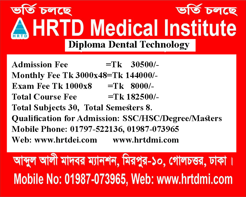
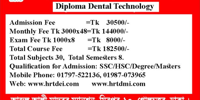
Dental Diploma Course Fee in Dhaka. MobileN No. 01987-073965, 01797-522136. Diploma in Dental 4 Years Tk 182500/-, Diploma in Dental Technology 4 Years Tk 182500/-, Diploma in Dental Technology 3 Years Tk 142500/-, Diploma in Dental Assistant 2 Years Tk 92500/-, Diploma in Dental Technology 2 Years Tk 92500/-, Dental Training Course 1 Year Tk 52500/-.
Dental Diploma Course Admission Fee in Dhaka
Dental Diploma Course Admission Fee in Dhaka. Mobile No. 01987-073965, 01797-522136. Diploma in Dental 4 Years Admission Fee Tk 30500/-, Diploma in Dental Technology 4 Years Admission Fee Tk 30500/-, Diploma in Dental Technology 3 Years Admission Fee Tk 20500/-, Diploma in Dental Assistant 2 Years Admission Fee Tk 16500/-, Diploma in Dental Technology 2 Years Tk 16500/-, Dental Training Course 1 Year Tk 12500/-. If you want to complete Dentist Best Diploma Course in Dhaka from HRTD Medical Institute, Please contact us.
Dentistry Diploma Course Location in Dhaka
Dentist Best Diploma Course Location in Dhaka. Mobile No. 01987-073965, 01797-522136. HRTD Medical Institute, Abdul Ali Madbor Mansion, Section-6, Block-Kha, Road-1, Plot-11, Mirpur-10 Golchattar, Metro Rail Piller No. 249, Dhaka-1216.
Dentistry Diploma Course Payment System
Dentistry Diploma Course Payment System. Mobile No. 01987-073965, 01797-522136.
Admission Fee + Monthly Fee + Exam Fee. Others Cost- Semester-wise Lecture Sheet+Book.
Dentistry Diploma Course in Dhaka Admission Eligibility
Dentistry Diploma Course in Dhaka Admission Eligibility. Mobile No. 01987-073965, 01797-522136. SSC or Equivalent/ HSC/Degree/ Masters from Any Background ( Science/ Arts/ Commerce/ Technical).
Some Subjects for Dentistry Diploma Course in Dhaka, Bangladesh
Dentistry Diploma Course Subjects. Mobile No. 01987-073965, 01797-522136.
- Human Anatomy & Physiology
- Dental Anatomy
- Dental Pharmacology
- Dental Surgery-1
- Dental Surgery-2
- Medical Microbiology,
- Hematology
- General Pathology
- Dental Pathology
- Dental Abscess & Treatment
- Dental Filling Materials
- Oral Surgery
- Oral Ulcer
- Clinical Practice In Dental Surgery
- Dental Sterilization
Admission System of Dentistry Diploma Course in Dhaka
অনলাইন এবং অফলাইন দুই সিস্টেমেই ভর্তির সুযোগ আছে।
Teachers for Dentistry Diploma Course in Dhaka
Dr. Sanjana, BDS
Dr. Nazmun Nahar Juthi, BDS
Dr. Sanjana Bite Ahmed, BDS, MPH
Dr. Sakulur Rahman, MBBS, CCD (BIRDEM)
Dr. Nurunnahar Keya, BDS,
Dr. Nazmun Nahar Juthi, BDS, PGT
Dr. Kamrunnahar Keya, BDS, PGT
Dr. Suhana, MBBS, PGT
Dr. Layla, MBBS, PGT
Dr. Lamia, MBBS
Dr. Disha, MBBS, FCPS (FP)
Dr. Afrin, MBBS, PGT
Dr. Tisha, MBBS, PGT
Dr. Danial Haque, MBBS, CCD
Dr. Turzo, MBBS, FCPS (FP)
Dr. Benzir, MBBS, FCPS (FP)
Dr. Trisha, MBBS, PGT
Dr. Farhana, MBBS, PGT
Dr. Jannatul Aman, MBBS, PGT
Dr. Mahinul Islam, MBBS
Dr. Amena Afroze Anu, MBBS, PGT
Class Time for Dentistry Diploma Course in Dhaka
Weekly Class 3 hours. For Regular Students Friday 1 hour, Saturday 1 hour, and Monday 1 hour. For Job holders, Friday is 3 hours, or Monday is 3 hours. Morning Shift 9:00 am to 12:00 pm, and Evening Shift 3:00 pm to 6:00 pm.
Dental Courses of HRTD Medical Institute
Dental Courses of HRTD Medical Institute. Mobile Phone 01797522136, 01987073965. Dental Training Course, Diploma Dental Assistant, Diploma in Dental Technology Course, Diploma in Dental, Post Diploma in Dental Surgery.
These Courses are helpful and essential for taking a job in Dental Hospitals, Health Service Centers, Dental Care Centers, Dental Services Centers, Health Sectors of NGOs, Health Centers of Schools, Health Centers of Colleges, Health Sectors of Universities, and Health Sectors of Garments Manufacturing Companies, Dental Research and Manufacturing Company, Dental Device Manufacturing Companies, Dental Chemical Manufacturing Companies, etc.
These Courses are also helpful for conducting a Dental Chamber, Dental Clinic, Dental Nursing Home, General Hospital, Medical College, Medical Institute, Health Research Institute, Medical Research Institute, Dental Research Institute, etc.
What is Dental Filling?
A dental filling is the fill-up of a small dental hole or cavity. To repair a cavity, a dentist removes the decayed tooth tissue and then fills the space with a filling material. The filling materials are of various types. Some are long-lasting and some are shot-lasting.
Causes of dental filling
When decay-causing bacteria come into contact with sugars and starches from foods and drinks, they form an acid. This acid can attack the tooth’s surface (enamel), causing it to lose minerals.
When a tooth is repeatedly exposed to acid, such as when you frequently consume food or drink high in sugar and starches, the enamel continues to lose minerals. A white spot may appear where minerals have been lost. This is a sign of early decay.
Tooth decay can be stopped or reversed at this point. Enamel can repair itself by using minerals from saliva and fluoride from toothpaste or through the application of fluoride by a dentist or dental hygienist. If more minerals are lost than can be restored, the enamel weakens and eventually breaks down, forming a cavity.
More severe decay can cause a large hole or even destruction of the entire tooth. If tooth decay is not treated, it can cause pain, infection, and even tooth loss. So dental filling treatment is very essential for a cavity patient.
What are the 5 dental problems?
Dentistry is one of the oldest fields of medicine, and it is vitally important to our overall health and well-being. That said, it is also one of the most commonly neglected aspects of health care, with many people failing to receive regular dental checkups and cleanings.
Unfortunately, this opens the door to a wide range of dental problems, all of which can have serious consequences if left untreated. Here are five of the most common dental problems:
1. Tooth Decay: Tooth decay is one of the most common dental problems and is caused by a buildup of plaque and bacteria on the teeth. Over time, this buildup can lead to cavities and tooth decay, which can cause serious pain and discomfort.
2. Gum Disease: Gum disease is caused by bacteria and plaque that accumulates at the gum line. If left untreated, it can lead to gum recession, tooth loss, and even bone loss.
3. Tooth Sensitivity: Tooth sensitivity is caused by a thinning of the enamel, which is the outer layer of the tooth. This can lead to pain when consuming hot or cold foods and drinks.
4. Tooth Abrasion: Tooth abrasion is caused by improper brushing techniques and can lead to the erosion of the enamel. Over time, this can cause the teeth to become more sensitive and can even lead to tooth loss.
5. Malocclusion: Malocclusion is the misalignment of the teeth, either due to genetics or other factors. This can lead to difficulty speaking and eating, as well as a higher risk of tooth decay and gum disease.
These five dental problems are just some of the many that can occur if proper oral hygiene is not maintained. Regular visits to the dentist for checkups and cleanings can help to prevent and address these issues and is essential for maintaining a healthy smile.
Subject Details for Dentistry Diploma Course in Dhaka
Human Anatomy and Physiology for Dentistry Diploma Course in Dhaka
The study of the Body Structure and its functions is Anatomy and Physiology. Here we discuss the systems of the Human Body and its Organs, Tissues, and Cells. The systems of the Human Body are the Digestive System, Respiratory System, Cardiovascular System, Skeletal System, Muscular System, Nervous System, Endocrine System, Immune System, Lymphatic System, Integumentary System, and Urinary System.
Pharmacology-1 for Dentistry Diploma Course in Dhaka
The study of Drugs and Medicine is called Pharmacology. Here we discuss group-wise drugs and their medicines in Pharmacology-1. Common Groups of Drugs are Pain Killer Drugs, Anti Ulcer Drugs, Anti Vomiting Drugs, Laxative Drugs, Motility Drugs, Antimotility Drugs, Bronchodilator Drugs, Antibiotic Drugs, Anti Fungal Drugs, Anti Protozoal Drugs, Anti Viral Drugs, Anthelmintic Drugs, Anti Hypertensive Drugs, Beta Blocker Drugs, Calcium Channel Blocker Drugs, ACE Inhibitor Drugs, Hemostatic Drugs, Analgesic Drugs, Antipyretic Drugs, Anti Thrombotic Drugs, etc.
Study of OTC Drugs for Dentistry Diploma Course in Dhaka
OTC Drugs are important for Diploma in Dental Course, Diploma Dental Assistant Course, all Medical Assistant Courses, Diploma Medical Courses, LMAF Courses, and RMP Courses. It is also important for the Diploma in Medicine & Diploma in Surgery Course. These OTC Drugs can be sold or purchased without any prescription from Registered MBBS Doctors. These Drugs are Emergency and Safe for the patients. The study of OTC Drugs improves the quality of practice. Some OTC Drugs are Albendazole, Ascorbic Acid, Calcium, Multivitamins, Vitamin B Complex, Omeprazole, Oral Rehydration Salt, Salbutamol, etc.
First Aid for Dentistry Diploma Course in Dhaka
First Aid is an important subject for Medical Courses including Diploma in Dental, Diploma in Medicine& Surgery Course, RMP Courses, LMAF Courses, Paramedical Courses, DMA Courses, DMS Courses, Nursing Courses, Dental Courses, Pathology Courses, Physiotherapy Courses, Caregiver Courses, etc. Here we discuss Shock, Classification Shock, Causes of Shock, Stages of Shock, Clinical Features of Shock, Hypovolemic Shock, Cardiogenic Shock, Neurogenic Shock, Traumatic Shock, Burn Shock, Electric Shock, Psychogenic Shock, Anaphylactic Shock, First Aid of Shock, First Aid of Cut, First of Snake Bite, First Aid of Accidental Injury, etc.
Hematology and Pathology for Dentistry Diploma Course in Dhaka
The study of Blood and Blood Disease is called Hematology and the Study of Pathos and Process of Disease Creation and Diagnosis is called Pathology. In Hematology and Pathology, we discuss blood cells, their structure and functions, Blood Diseases, Common Pathos and their pathogenesis, Atrophy, Hypertrophy, Metaplasia, Gangrene, Pathological Tests like TC, DC, ESR, Hemoglobin Percentage,
Microbiology and Antimicrobial Drugs for Dentistry Diploma Course in Dhaka
The Study of Microorganisms is called Microbiology. The Drugs that are used for the treatment of Infectious Diseases are Antimicrobial Drugs. Microorganisms are Bacteria, Protozoa, Fungus, and Virus. Antimicrobial Drugs are Antibiotic Drugs ( Antibacterial Drugs), Anti Protozoal Drugs, Anti Fungal Drugs, and Anti Viral Drugs. Antibacterial Drugs are Azithromycin, Erythromycin, Clarithromycin, Cefaclor, Cefixime, Cefuroxime, Ceftriaxone, Ciprofloxacin, Moxifloxacin, Doxicicline, Gentamycin, Neomycin, Flucloxacillin, Amoxicillin, Clindamycin, etc. Antiprotozoal Drugs are Metronidazole, Secnidazole, Tinidazole, Ornidazole, Nitazoxanide, etc. Antifungal Drugs are Fluconazole, Ketoconazole, Itraconazole, Econazole, Miconazole, Terbinafine, etc.
Cardiovascular Anatomy & Physiology for Dentistry Diploma Course in Dhaka
Cardiovascular Anatomy is a branch of Anatomy, and Cardiovascular Physiology is a branch of Physiology. These two subjects are related to cardiology. Cardiovascular Anatomy and Physiology are being studied in a single subject for Diploma Medical Courses, Paramedical Courses, and All Short Medical Courses.
We discuss here the Anatomy of the Heart, Cardiac Chambers, Cardiac Valves, Cardiac Wall, Cardiac Septum, Right Heart, Left Heat, Function of Right Heat, Functions of Left Heart, Aorta, Venecava, Artery, Vein, Capillary, Pulmonary Blood Circulation, Cerebral Blood Circulation, Renal Blood Circulation, Hepatic Blood Circulation, Portal Vein and Portal Circulation, Heart Beat, Pulse, Pulse Rate, Tachycardia, Bradycardia, Blood Pressure, Normal Blood Pressure, Hypertension, Hypotension, Stroke Volume, Cardiac Output, Heart Failure, etc. This Subject is the most essential for the Diploma in Medicine and Diploma in Surgery Course.
Practice of Medicine for Dentistry Diploma Course in Dhaka
The study of Disease and Treatment is called the Practice of Medicine. This subject is important for Diploma in Dental, Dental Practitioners, Diploma Medical Practitioner, Diploma Medical Assistant, and Rural Medical Practitioner. This subject discusses some common diseases. The discussion points for the Practice of Medicine are the Definition of Disease, Causes of Disease, Clinical Features of Disease ( Symptoms and Signs), Investigation of Disease, Treatment of Disease, Complication of Disease, and Advice for the Patients.
Dental Anatomy & Physiology for Dentistry Diploma Course in Dhaka
Dental Anatomy refers to the study of the structure, form, and development of the teeth and surrounding oral tissues. It includes understanding tooth morphology, types of teeth (incisors, canines, premolars, molars), their functions, and how they fit together in the dental arch.
Dental Physiology focuses on the functions and processes of the oral cavity, including tooth development (odontogenesis), chewing (mastication), saliva production, and the role of teeth in speech and digestion.
Anatomy of Headneck for Dentistry Diploma Course in Dhaka
The anatomy of the head and neck is complex, comprising bones, muscles, nerves, blood vessels, glands, and other structures essential for vital functions like respiration, circulation, and sensory perception. Here’s a breakdown:
1. Bones of the Head and Neck
- Skull: Protects the brain and supports facial structures. It has two main parts:
- Cranium: Encloses the brain (frontal, parietal, temporal, occipital, sphenoid, and ethmoid bones).
- Facial bones: Form the face (maxilla, mandible, nasal bones, zygomatic, etc.).
- Hyoid Bone: U-shaped bone in the neck supporting the tongue and swallowing muscles.
- Cervical Vertebrae: Seven vertebrae (C1-C7) forming the neck, including:
- Atlas (C1): Supports the skull.
- Axis (C2): Allows head rotation.
💪 2. Muscles of the Head and Neck
- Facial Muscles: Control facial expressions (e.g., orbicularis oculi, orbicularis oris).
- Masticatory Muscles: Aid chewing (e.g., masseter, temporalis).
- Neck Muscles: Support head movement and swallowing:
- Sternocleidomastoid: Turns and flexes the head.
- Trapezius: Moves the shoulder and extends the neck.
- Suprahyoid & Infrahyoid Muscles: Assist in swallowing.
🩸 3. Blood Supply and Venous Drainage
- Arteries:
- Common Carotid Arteries: Main supply to the head and neck, bifurcating into:
- Internal Carotid: Brain and eyes.
- External Carotid: Face, scalp, and neck.
- Common Carotid Arteries: Main supply to the head and neck, bifurcating into:
- Veins:
- Jugular Veins: Drain blood from the head and neck into the heart.
📊 4. Nerves of the Head and Neck
- Cranial Nerves (12 pairs): Key for sensory and motor functions.
- Notable ones include:
- Trigeminal (V): Sensation to the face, motor to chewing muscles.
- Facial (VII): Controls facial muscles and taste.
- Vagus (X): Innervates heart, lungs, and digestive tract.
- Notable ones include:
- Cervical Plexus: Nerves from C1-C4 supply sensation and motor function to the neck.
🧬 5. Glands and Lymphatics
- Salivary Glands: Produce saliva (parotid, submandibular, sublingual).
- Thyroid and Parathyroid Glands: Regulate metabolism and calcium.
- Lymph Nodes: Filter lymphatic fluid and are located throughout the neck.
👁️ 6. Special Sensory Structures
- Eyes: Vision and protected by the bony orbit.
- Ears: Hearing and balance.
- Nose: Olfaction (smell) and respiratory passage.
- Mouth: Speech, digestion, and taste.
Neuro Anatomy for Dentistry Diploma Course in Dhaka
Neuroanatomy is the study of the structure and organization of the nervous system. It is divided into the central nervous system (CNS) and the peripheral nervous system (PNS).
🧠 1. Central Nervous System (CNS)
The CNS includes the brain and spinal cord, responsible for processing and integrating information.
A. Brain
The brain is divided into major regions, each with specialized functions:
a) Cerebrum
- Largest part of the brain, responsible for higher cognitive functions.
- Divided into two hemispheres (right and left) connected by the corpus callosum.
- Lobes of the Cerebrum:
- Frontal Lobe: Motor control, speech (Broca’s area), decision-making, and personality.
- Parietal Lobe: Sensory perception (touch, temperature, pain), spatial awareness.
- Temporal Lobe: Hearing, memory, and language comprehension (Wernicke’s area).
- Occipital Lobe: Vision processing.
b) Diencephalon
Located deep within the brain, it includes:
- Thalamus: Sensory relay center.
- Hypothalamus: Regulates hormones, hunger, thirst, and body temperature.
- Epithalamus: Includes the pineal gland, which regulates circadian rhythms.
c) Brainstem
Connects the brain to the spinal cord, controlling vital functions.
- Midbrain: Visual and auditory reflexes.
- Pons: Relays information between cerebrum and cerebellum; regulates breathing.
- Medulla Oblongata: Controls autonomic functions (heart rate, respiration, digestion).
d) Cerebellum
Located at the back of the brain, it coordinates balance, posture, and fine motor movements.
B. Spinal Cord
- Structure: Cylindrical structure extending from the medulla to the lower back.
- Function: Transmits sensory and motor information between the brain and body.
- Segments: Cervical, thoracic, lumbar, sacral, and coccygeal regions.
- Gray Matter: Contains nerve cell bodies (butterfly-shaped in cross-section).
- White Matter: Contains myelinated nerve fibers for communication.
🔍 2. Peripheral Nervous System (PNS)
The PNS connects the CNS to the body and consists of nerves and ganglia.
A. Cranial Nerves (12 Pairs)
Emerge from the brainstem and control sensory and motor functions in the head and neck. Key examples:
- Optic (II): Vision.
- Trigeminal (V): Facial sensation, chewing.
- Vagus (X): Parasympathetic control of heart, lungs, and digestion.
B. Spinal Nerves (31 Pairs)
Arise from the spinal cord and carry sensory, motor, and autonomic signals.
- Cervical (8), Thoracic (12), Lumbar (5), Sacral (5), Coccygeal (1).
C. Autonomic Nervous System (ANS)
Controls involuntary functions (heart rate, digestion).
- Sympathetic: “Fight or flight” response (increases heart rate, dilates pupils).
- Parasympathetic: “Rest and digest” response (slows heart rate, promotes digestion).
🧬 3. Functional Pathways
- Sensory (Afferent): Carries sensory information from the body to the CNS.
- Motor (Efferent): Transmits motor commands from the CNS to muscles.
- Reflex Arc: Simple neural circuit allowing rapid, automatic responses.
📊 4. Meninges and Cerebrospinal Fluid (CSF)
- Meninges: Protective layers surrounding the CNS:
- Dura Mater: Tough outer layer.
- Arachnoid Mater: Middle layer with CSF circulation.
- Pia Mater: Delicate inner layer on the brain surface.
- CSF: Cushions the brain and spinal cord, circulating in the subarachnoid space.
📡 5. Blood Supply to the Brain
- Circle of Willis: A vital arterial network supplying the brain.
- Internal Carotid Arteries and Vertebral Arteries: Main blood suppliers.
Clinical Practice in Dental Surgery for Dentistry Diploma Course in Dhaka
🦷 Clinical Practice in Dental Surgery
Clinical practice in dental surgery involves diagnosing, treating, and preventing diseases, injuries, and conditions affecting the teeth, gums, jaws, and oral tissues. It combines theoretical knowledge, clinical skills, patient management, and surgical expertise.
Patient Assessment and Diagnosis
History Taking
- Medical History: Systemic conditions (e.g., diabetes, cardiovascular disease), medications, allergies.
- Dental History: Previous dental treatments, pain history, oral hygiene practices.
- Social History: Smoking, alcohol, lifestyle, and occupational factors affecting oral health.
Clinical Examination
- Extraoral: Inspect face, jaw, temporomandibular joint (TMJ), and lymph nodes.
- Intraoral: Evaluate teeth, gums, oral mucosa, tongue, palate, and occlusion.
Diagnostic Tools
- Radiographs (X-rays): Periapical, panoramic (OPG), cone-beam CT (CBCT).
- Vitality Testing: Assess pulp health using cold, heat, or electric tests.
- Impressions and Models: For treatment planning (e.g., prosthetics, orthodontics).
Dental surgery ranges from minor procedures to complex oral surgeries.
Tooth Extraction
- Indications: Caries, periodontal disease, impacted teeth, orthodontic reasons.
- Techniques:
- Simple Extraction: For visible, easily accessible teeth.
- Surgical Extraction: For impacted, fractured, or difficult teeth (e.g., wisdom teeth)
Impacted Tooth Removal
- Common in Third Molars (Wisdom Teeth): Requires surgical exposure and sectioning.
Anesthesia and Pain Management
- Local Anesthesia: Lidocaine, articaine—common for routine dental surgery.
- Sedation: Nitrous oxide (inhalation), IV sedation for anxious patients.
- General Anesthesia: For complex oral and maxillofacial surgeries.
Oral & Dental Pathology for Dentistry Diploma Course in Dhaka
There are many acquired and inherited developmental abnormalities that alter the size, shape and number of teeth.
Supernumerary teeth- Presence of extra. Such as-extra incisor, extra molar.
Cleidocranial dysplasia- ৫০ এর অধিক দাঁত হলে তাকে Cleidocranial dysplasia বলে। ইহা একটি autosomal dominant disorder.
Oligodontia-জন্মগতভাবে ৬ বা অধিক দাঁতের মিছিং থাকলে তাকে ওলিগডনশিয়া বলে।
Taurodontism-টরোডন্টিজম হচ্ছে দাঁতের একটি গঠনগত অস্বাভাবিকতা ।
Fusion-দুটি সংলগ্ন দাঁতের জীবাণু একত্রিত হয়ে একটি বড় দাঁত তৈরি করে। দাঁতের ফিউশনের ঘটনাটি
Oral Ulcer for Dentistry Diploma Course in Dhaka
Oral ulcers are painful sores or lesions that occur on the mucous membrane inside the mouth. They can result from trauma, infection, systemic diseases, or other underlying conditions.
Types of Oral Ulcers
Oral ulcers are classified based on their duration, cause, and appearance:
a) Traumatic Ulcers
- Cause: Mechanical injury (e.g., sharp teeth, dental appliances), chemical burns, or thermal injury.
- Features: Single ulcer with irregular margins, surrounded by inflamed tissue.
- Healing Time: 1-2 weeks after removing the cause.
b) Aphthous Ulcers (Canker Sores)
- Cause: Unknown (possible immune dysfunction, stress, hormonal changes, or nutritional deficiency).
- Types:
- Minor: Small (<1 cm), shallow, round, heals in 1-2 weeks.
- Major: Larger (>1 cm), deeper, can last 4-6 weeks, may scar.
- Herpetiform: Multiple small (<3 mm) clustered ulcers, heals in 1-2 weeks.
c) Infectious Ulcers
- Viral:
- Herpes Simplex Virus (HSV): Primary herpetic gingivostomatitis, recurrent herpes labialis (cold sores).
- Varicella-Zoster Virus: Oral lesions in shingles.
- Hand-Foot-Mouth Disease: Coxsackievirus.
- Bacterial: Secondary to trauma or poor oral hygiene (e.g., syphilis, tuberculosis).
- Fungal: Oral candidiasis-related ulcers (in immunocompromised patients).
d) Systemic Disease-Related Ulcers
- Behçet’s Disease: Recurrent oral ulcers with genital ulcers and eye inflammation.
- Lichen Planus: Chronic inflammatory condition with white striations and ulcers.
- Lupus Erythematosus: Autoimmune disease with discoid oral ulcers.
- Inflammatory Bowel Disease (IBD): Crohn’s disease and ulcerative colitis.
e) Drug-Induced Ulcers
- Causes: Chemotherapy, NSAIDs, beta-blockers.
- Presentation: Painful, non-healing ulcers.
f) Malignant Ulcers
- Cause: Oral squamous cell carcinoma.
- Features: Non-healing ulcer with indurated (hardened) edges, pain, and bleeding. Requires biopsy for confirmation.
Clinical Features of Oral Ulcers
- Shape: Round or oval.
- Size: Small (1-2 mm) to large (>1 cm).
- Color: Yellow/white base with erythematous (red) border.
- Pain: Ranges from mild discomfort to severe pain.
- Location: Buccal mucosa, tongue, soft palate, gingiva.
Clinical History
- Duration, recurrence, trauma, systemic symptoms, medication use.
- Risk factors: Smoking, alcohol use, immunosuppression.
b) Clinical Examination
- Number, size, location, and appearance of ulcers.
- Check for associated lymphadenopathy (swollen lymph nodes).
c) Investigations
- Biopsy: For non-healing or suspicious ulcers (rule out malignancy).
- Blood Tests:
- Complete blood count (CBC) for anemia.
- Autoimmune markers (ANA, anti-dsDNA).
- Viral serology (HSV, HIV).
When to Refer
- Non-healing ulcers lasting >3 weeks.
- Suspected malignancy (indurated, persistent, or ulcerated lesions).
- Multiple ulcers with systemic involvement (e.g., Behçet’s).
- Recurrent or severe ulcers unresponsive to treatment.
Dental Filling Materials for Dentistry Diploma Course in Dhaka
Dental Filling Materials
Dental filling materials are used to restore the function, integrity, and structure of teeth damaged by caries (tooth decay), trauma, or wear. Different materials offer unique properties suited to specific clinical situations.
 HRTD Medical Institute


One comment
Pingback: Best Diploma in Public Health 1 year | HRTD Medical Institute